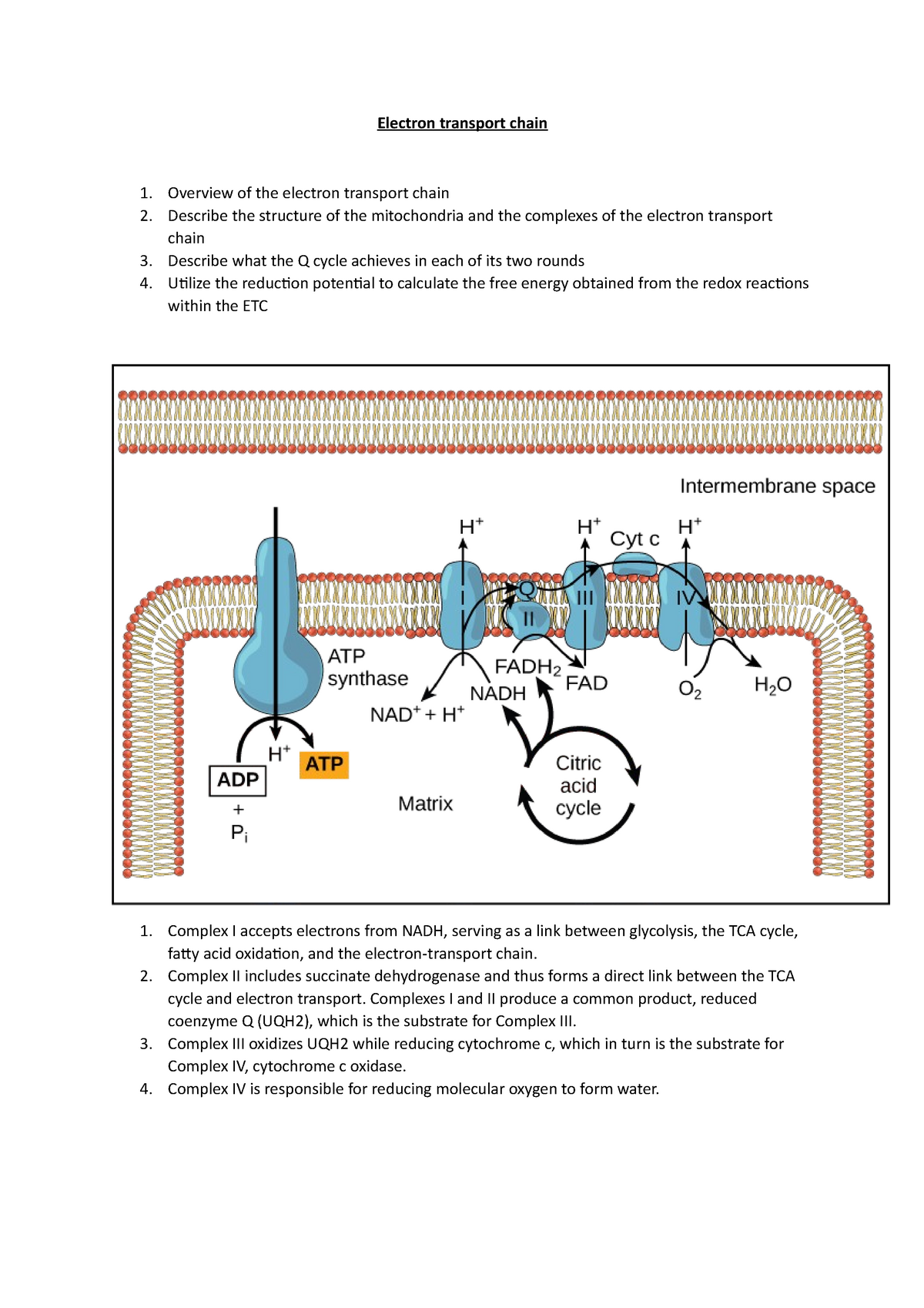
The Succinate Dehydrogenase (SDH) complex, also known as mitochondrial Complex II, is a unique enzymatic powerhouse that serves as a vital functional bridge between the Citric Acid Cycle (TCA cycle) and the Electron Transport Chain (ETC). By facilitating the oxidation of succinate to fumarate while simultaneously reducing ubiquinone to ubiquinol, it plays a non-redundant role in cellular energy production and metabolic signaling.
1. The Dual Role of Complex II in Cellular Metabolism
Unlike any other enzyme in the mitochondrial landscape, the succinate dehydrogenase complex 2 holds a dual residency. It is an integral member of the Citric Acid Cycle (Krebs cycle) and a fundamental component of the respiratory chain. This dual functionality is not merely a biological curiosity but a strategic evolutionary adaptation for metabolic efficiency.
In the Citric Acid Cycle, SDH catalyzes the sixth step: the oxidation of succinate into fumarate. During this reaction, two hydrogen atoms are removed from succinate. Unlike other steps in the cycle that reduce $NAD^+$ to $NADH$, the redox potential of the succinate/fumarate couple is not high enough to reduce $NAD^+$. Therefore, SDH utilizes a covalently bound Flavin Adenine Dinucleotide (FAD) cofactor as the initial electron acceptor.
Once the electrons are captured by FAD, the complex transitions into its role as Complex II of the Electron Transport Chain. It funnels these electrons through a series of iron-sulfur (Fe-S) clusters directly to the ubiquinone (Coenzyme Q) pool in the inner mitochondrial membrane. By bypassing Complex I, SDH provides an alternative entry point for electrons into the respiratory chain, ensuring that energy production can continue even if NADH levels are low or if Complex I is inhibited. As of 2026, this "metabolic bypass" remains a primary focus for researchers studying cellular resilience under oxidative stress.
2. Structural Composition and the Four Essential Subunits
The architectural integrity of the succinate dehydrogenase complex 2 is maintained by four distinct subunits, encoded by nuclear DNA (SDHA, SDHB, SDHC, and SDHD). Understanding the specific roles of these subunits is crucial for diagnosing the various mitochondrial and oncogenic pathologies associated with the complex.
- SDHA (The Flavoprotein): This is the catalytic heart of the complex. It contains the FAD binding site where succinate is converted to fumarate. SDHA is a hydrophilic protein that extends into the mitochondrial matrix, providing the necessary surface area for substrate interaction.
- SDHB (The Iron-Sulfur Protein): This subunit acts as the electrical wire of the complex. It houses three distinct iron-sulfur clusters—[2Fe-2S], [4Fe-4S], and [3Fe-4S]. These clusters facilitate the rapid "tunneling" of electrons from the FAD in SDHA toward the membrane-bound subunits.
- SDHC and SDHD (The Hydrophobic Anchors): These two subunits are integral membrane proteins that anchor the SDHA/SDHB catalytic dimer to the inner mitochondrial membrane. Together, they form the cytochrome b portion of the complex and contain the binding site for ubiquinone (the Q-site). They also house a heme b group, which, while not directly involved in the primary electron path to ubiquinone in all species, is believed to enhance the structural stability of the complex and may play a role in preventing the leakage of electrons.
The assembly of these four subunits is a tightly regulated process requiring specific assembly factors (SDHAF1, SDHAF2, SDHAF3, and SDHAF4). Any defect in these assembly proteins can lead to a functional deficiency of Complex II, even if the primary subunit genes are healthy.
3. The Mechanics of Electron Transfer and the Q-Site
The primary task of the succinate dehydrogenase complex 2 is the precise movement of electrons. This process begins at the dicarboxylate binding site in SDHA. When succinate binds, it transfers two electrons to the FAD, reducing it to $FADH_2$. However, the respiratory chain operates on single-electron transfers.
To manage this, the electrons are passed one by one through the Fe-S clusters in SDHB. The spatial arrangement of these clusters is optimized for quantum tunneling, allowing electrons to move across relatively large molecular distances with minimal energy loss. The final destination for these electrons is the ubiquinone binding site (Q-site), located in the cleft between SDHB, SDHC, and SDHD.
There are actually two potential Q-sites: $Q_P$ (proximal to the matrix) and $Q_D$ (distal). Most physiological electron transfer occurs at the $Q_P$ site. When ubiquinone (Q) accepts two electrons and two protons from the matrix, it is reduced to ubiquinol ($QH_2$). This ubiquinol then diffuses through the lipid bilayer to Complex III. A critical aspect of this mechanism is the prevention of electron "slippage." If electrons escape the pathway before reaching ubiquinone, they can react with molecular oxygen to form reactive oxygen species (ROS), such as superoxide. Under normal 2026 health conditions, SDH is remarkably efficient, but mutations in the Q-site are a well-known cause of increased oxidative stress.
4. Why Complex II Does Not Pump Protons
A defining characteristic of the succinate dehydrogenase complex 2—and one that distinguishes it from Complexes I, III, and IV—is that it does not contribute directly to the proton gradient across the inner mitochondrial membrane. It does not "pump" protons from the matrix into the intermembrane space.
The reason for this lies in the thermodynamics of the reaction. The change in Gibbs free energy ($\Delta G$) associated with the transfer of electrons from succinate to ubiquinone is relatively small. In Complexes I, III, and IV, the redox reactions release enough energy to drive the conformational changes required to move protons against their concentration gradient. For Complex II, the energy released is just enough to facilitate electron transfer but insufficient to power the molecular machinery of a proton pump.
Consequently, Complex II is often considered less "efficient" in terms of ATP yield per electron pair compared to Complex I. While the oxidation of NADH (via Complex I) leads to the pumping of 10 protons, the oxidation of succinate (via Complex II) only leads to the pumping of 6 protons (occurring later at Complexes III and IV). Despite this, Complex II is essential because it allows the cell to utilize succinate, a major metabolic intermediate, as a direct fuel source for the respiratory chain.
5. Regulation via Post-Translational Modifications
As of 2026, scientific understanding of how the succinate dehydrogenase complex 2 is regulated has expanded significantly beyond simple substrate availability. The complex is heavily regulated by post-translational modifications (PTMs), which allow the cell to fine-tune energy production based on current demands.
- Acetylation: SDHA is subject to reversible acetylation. High levels of acetylation generally inhibit the activity of the complex. The mitochondrial deacetylase SIRT3 is responsible for removing these acetyl groups, thereby activating SDH. This provides a direct link between the cell’s NAD+ levels (which SIRT3 requires) and the activity of the TCA cycle.
- Phosphorylation: Various kinases can phosphorylate the subunits of Complex II, affecting its assembly and its affinity for succinate. This allows for rapid signaling-based control of metabolism, often in response to growth factors or cellular stress.
- Competitive Inhibition: SDH is naturally inhibited by oxaloacetate, the end product of the TCA cycle. When oxaloacetate levels are high, it binds to the SDHA active site, slowing down the cycle to prevent the overproduction of intermediates. This feedback loop is essential for metabolic homeostasis.
| Feature | SDHA | SDHB | SDHC/SDHD |
|---|---|---|---|
| Location | Mitochondrial Matrix | Matrix Side / Membrane Interface | Inner Mitochondrial Membrane |
| Primary Cofactor | FAD (Covalently bound) | Three Fe-S Clusters | Heme b and Ubiquinone |
| Main Function | Succinate Oxidation | Electron Tunneling | Membrane Anchor & Q-reduction |
| Related Disease | Leigh Syndrome | Paraganglioma (PGL4) | Paraganglioma (PGL3 / PGL1) |
| Assembly Factors | SDHAF2 | SDHAF1, SDHAF3, SDHAF4 | N/A (Integrated during assembly) |
6. The "Oncometabolite" Connection and Cancer Pathology
Perhaps the most clinically significant insight into the succinate dehydrogenase complex 2 is its role as a tumor suppressor. Mutations in the genes encoding SDH subunits are directly linked to several types of cancer, most notably hereditary paragangliomas and pheochromocytomas (PGL/PCC), as well as certain gastrointestinal stromal tumors (GIST) and renal cell carcinomas.
When SDH is dysfunctional due to a mutation, succinate begins to accumulate in the mitochondrial matrix and leaks into the cytosol. At high concentrations, succinate acts as an oncometabolite. It structurally resembles $\alpha$-ketoglutarate ($\alpha$-KG) and competitively inhibits $\alpha$-KG-dependent dioxygenases.
One critical target is Prolyl Hydroxylase (PHD). Under normal oxygen levels, PHD marks the Hypoxia-Inducible Factor ($\text{HIF}-1\alpha$) for degradation. However, when accumulated succinate inhibits PHD, $\text{HIF}-1\alpha$ is stabilized and moves to the nucleus, even in the presence of oxygen. This creates a state of pseudohypoxia, triggering the expression of genes involved in angiogenesis, glucose transport, and glycolysis—all of which promote tumor growth and survival. This discovery has revolutionized the 2026 approach to metabolic oncology, making SDH a primary target for genetic screening in patients with specific neuroendocrine tumors.
7. Diagnostic and Therapeutic Implications in 2026
The status of the succinate dehydrogenase complex 2 is now a standard biomarker in many clinical pathologies. Immunohistochemistry (IHC) for SDHB is frequently used in pathology labs to screen for SDH-deficient tumors. A loss of SDHB staining typically indicates a mutation in any of the four subunits (A, B, C, or D), as the entire complex usually destabilizes when one component is missing or defective.
Beyond oncology, SDH is a focal point in the study of neurodegenerative diseases. Deficiencies in Complex II activity have been observed in the striatum of patients with Huntington’s disease, suggesting that metabolic failure at this specific junction may contribute to neuronal death.
Therapeutically, researchers are exploring "succinate prodrugs" to bypass certain metabolic blocks and SDH inhibitors for specific agricultural and medicinal applications. In the context of ischemia-reperfusion injury, the accumulation of succinate during the ischemic phase and its rapid oxidation upon reperfusion is a major source of ROS-mediated damage. Strategies to modulate SDH activity during these transitions are currently under investigation to protect heart and brain tissue during surgery or stroke recovery.
Frequently Asked Questions
What is the difference between Complex I and Complex II?
Complex I (NADH dehydrogenase) oxidizes NADH and pumps four protons across the membrane per electron pair. Complex II (Succinate dehydrogenase) oxidizes succinate and does not pump any protons. Complex II provides a way for the respiratory chain to function independently of the NADH/NAD+ ratio.
How do mutations in SDH lead to Leigh Syndrome?
Leigh Syndrome is a severe neurological disorder usually appearing in infancy. While most commonly associated with Complex I or IV defects, mutations in SDHA can impair the energy production required for the developing brain, leading to the characteristic subcortical brain lesions and psychomotor regression associated with the disease.
Is succinate dehydrogenase the same as succinate-coenzyme Q reductase?
Yes, these are different names for the same enzyme complex (Complex II). "Succinate dehydrogenase" emphasizes its role in the TCA cycle (removing hydrogens from succinate), while "Succinate-coenzyme Q reductase" emphasizes its role in the ETC (transferring those electrons to Coenzyme Q).
Why is SDHB immunohistochemistry used for screening?
SDHB is the "linchpin" for the stability of the entire complex. If there is a germline or somatic mutation in SDHA, SDHB, SDHC, or SDHD, the complex often fails to assemble correctly, and the SDHB protein is degraded. Therefore, the absence of SDHB staining in a tumor sample is a highly reliable surrogate marker for a mutation in any SDH subunit.
Can diet or supplements affect Complex II function?
While general mitochondrial health is supported by cofactors like Riboflavin (precursor to FAD) and Iron (for Fe-S clusters), there is no specific supplement that "boosts" Complex II beyond physiological norms in healthy individuals. However, in specific genetic deficiencies, high-dose riboflavin therapy is sometimes utilized to stabilize the SDHA subunit.
Practical Recommendation for Clinicians and Researchers
When evaluating patients with early-onset hypertension or unusual neuroendocrine growths, prioritize genetic screening for the SDHx gene suite. Given the 2026 guidelines for precision medicine, identifying an SDH deficiency early can lead to life-saving surveillance for associated malignancies. For laboratory researchers, focusing on the SDH-HIF1$\alpha$ axis remains the most promising path for developing small-molecule inhibitors that could potentially reverse the pseudohypoxic state in SDH-deficient cancers.