Ameloblastoma treatment focuses on the complete surgical removal of the tumor to prevent recurrence, which often involves a segmental or marginal resection of the jawbone. Following resection, the focus shifts to microvascular reconstruction using bone grafts and 3D-planned implants to restore facial symmetry, speech, and masticatory function.
Ameloblastoma is a rare, slow-growing, but locally aggressive tumor that arises from the odontogenic epithelium—the cells responsible for tooth development. While it is classified as a benign neoplasm because it rarely metastasizes to distant organs, its tendency to invade local bone and soft tissue makes it a significant clinical challenge. Left untreated, an ameloblastoma can cause severe facial disfigurement and compromise essential functions like breathing, swallowing, and speaking.
As of 2026, the management of ameloblastoma has evolved from a purely surgical discipline into a multidisciplinary approach involving oral and maxillofacial surgeons, pathologists, microvascular reconstructive surgeons, and increasingly, molecular oncologists. This guide examines the contemporary standards for diagnosing, treating, and reconstructing the jaw following an ameloblastoma diagnosis.
Diagnostic Prerequisites and Pre-Surgical Planning
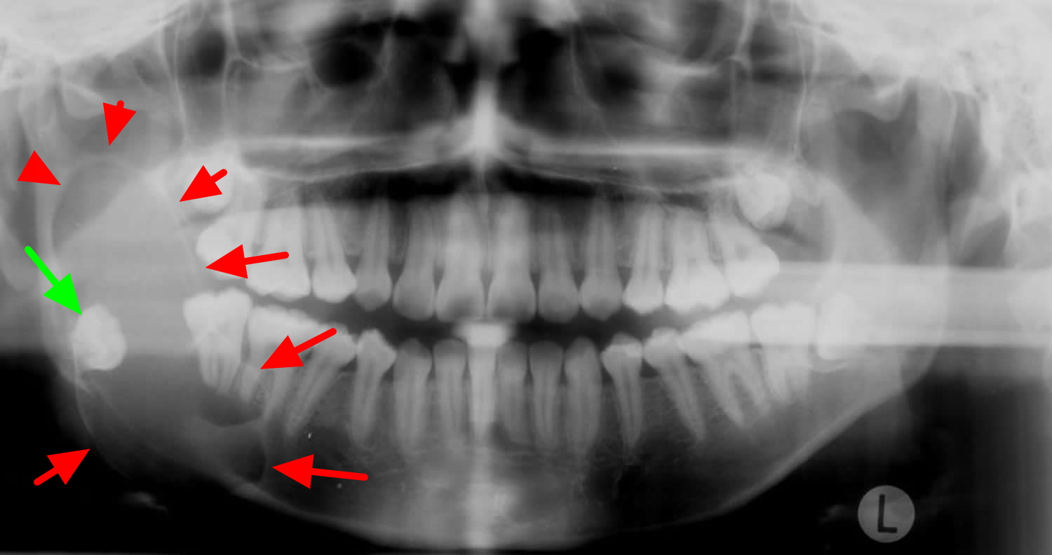
Before any treatment begins, a precise diagnosis is mandatory. Ameloblastomas are often discovered incidentally during routine dental X-rays, appearing as a "soap bubble" or "honeycomb" radiolucency. However, radiographic appearance alone is insufficient for planning radical surgery.
Histopathological Verification
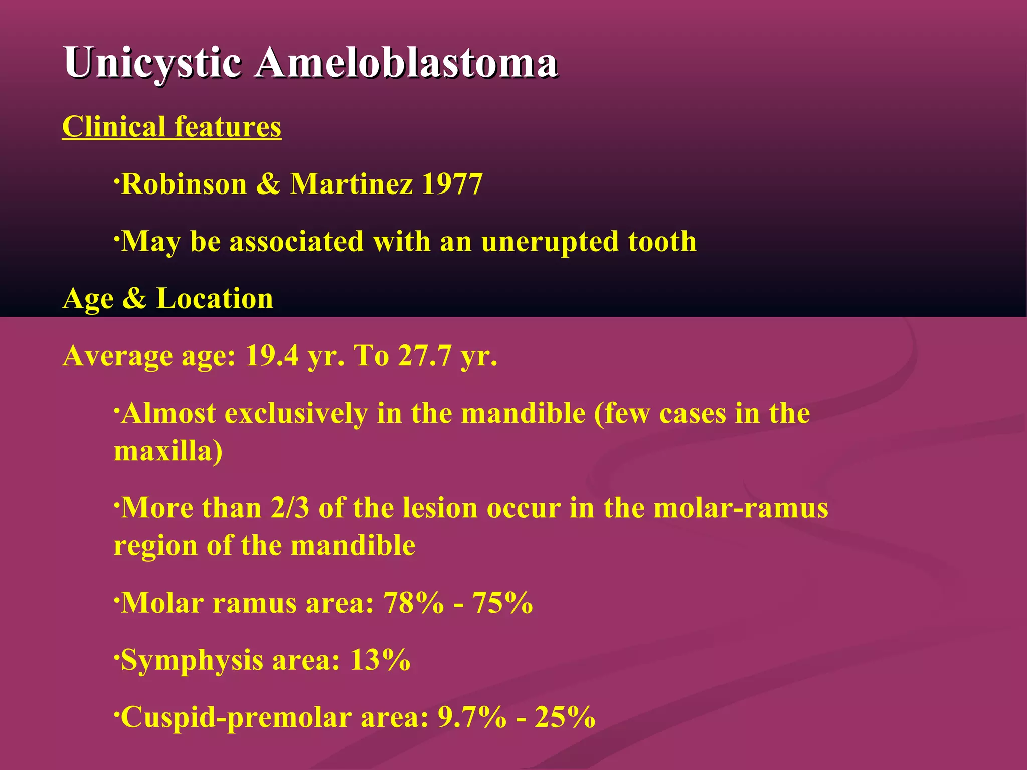
A tissue biopsy is the gold standard. Pathologists categorize the tumor into several types, most commonly the solid/multicystic type and the unicystic type. The solid type is the most aggressive and carries the highest risk of recurrence. In recent years, molecular testing for the BRAF V600E mutation has become a standard diagnostic step. Found in approximately 60% to 80% of mandibular ameloblastomas, this mutation not only confirms the diagnosis but also opens the door for targeted medical therapies if surgery is high-risk.
Advanced Imaging
In 2026, Cone Beam Computed Tomography (CBCT) and high-resolution MRI are the primary tools for mapping the tumor’s boundaries. While CT scans provide excellent detail of the bone destruction, MRI is superior for identifying "skip lesions" or soft tissue invasion into the masticatory spaces. Surgeons use these images to determine the necessary surgical margins—the amount of healthy bone that must be removed alongside the tumor to ensure no microscopic cells are left behind.
Surgical Treatment Modalities
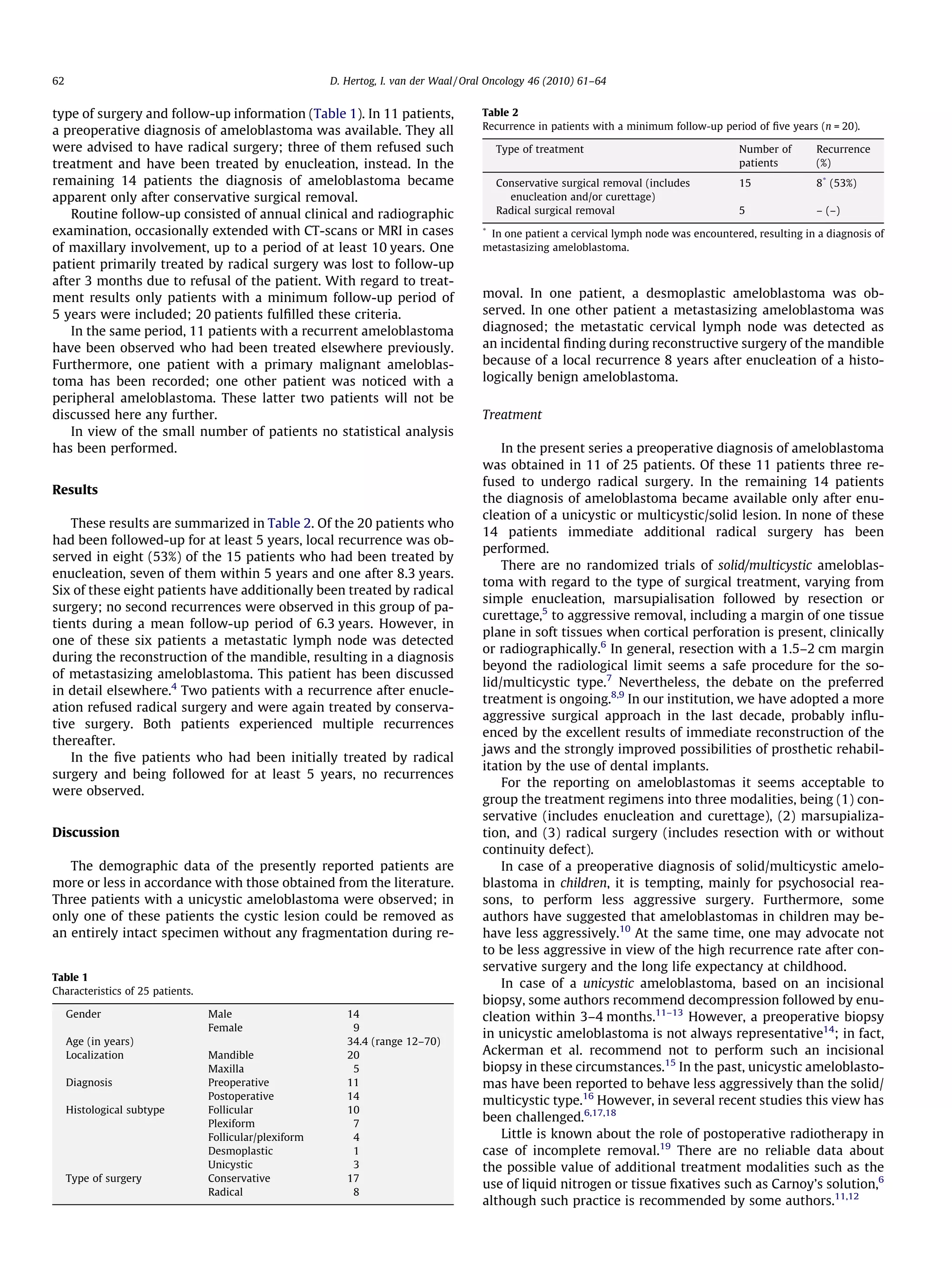
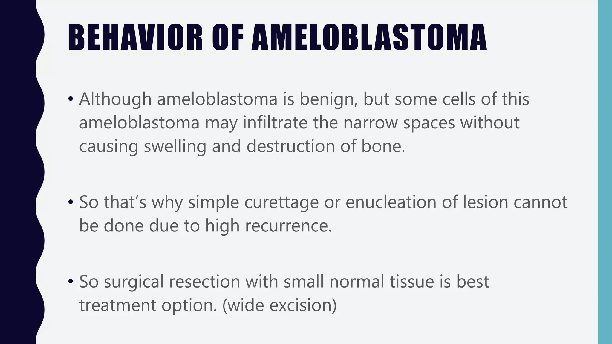
The primary goal of ameloblastoma treatment is the total eradication of the tumor. History has shown that "conservative" methods, while less invasive, often lead to a high rate of recurrence, sometimes decades after the initial procedure.
Radical Resection: The Gold Standard
For solid or multicystic ameloblastomas, radical resection is the most reliable treatment. This involves removing the tumor with a 1.0 to 1.5-centimeter margin of healthy-appearing bone.
- Marginal Resection: This technique removes the tumor while maintaining the continuity of the lower border of the mandible. It is only used when the tumor has not invaded the full thickness of the jawbone.
- Segmental Resection: This involves removing a complete section of the jaw. This is necessary when the tumor is large or has perforated the bone cortex. While more disfiguring initially, it has the lowest recurrence rate (less than 10%).
Conservative Management: Curettage and Enucleation
In specific cases of unicystic ameloblastoma—a less aggressive variant often found in younger patients—surgeons may opt for enucleation (scooping out the tumor) followed by curettage (scraping the bone walls). To further reduce recurrence, chemical cauterization using Carnoy’s solution or liquid nitrogen (cryotherapy) is often applied to the bone cavity. Despite these adjuncts, the recurrence rate for conservative treatment remains significantly higher than radical resection, necessitating lifelong follow-up.
The Role of Targeted Molecular Therapy
One of the most significant shifts in ameloblastoma treatment leading into 2026 is the use of BRAF inhibitors. For patients with large, unresectable tumors or those who are medically unfit for extensive surgery, drugs like Dabrafenib and Trametinib have shown the ability to shrink tumors significantly.
This approach, known as neoadjuvant therapy, can reduce the size of a massive tumor until it is small enough to be removed with a less invasive "marginal" resection rather than a "segmental" one. While not yet a replacement for surgery, molecular therapy is a critical tool for preserving jaw continuity in complex cases.
Jaw Reconstruction and Functional Restoration
Losing a segment of the jaw is a traumatic experience for a patient. Modern reconstruction techniques aim to restore both the "form" (how the face looks) and the "function" (how the patient eats and speaks) in a single surgical session, often referred to as "jaw-in-a-day."
Virtual Surgical Planning (VSP)
In 2026, surgery is rarely performed without Virtual Surgical Planning. Using 3D models generated from the patient's CT scans, surgeons "perform" the operation on a computer first. They design custom cutting guides and titanium plates that are 3D-printed to fit the patient's unique anatomy perfectly. This precision reduces time under anesthesia and improves aesthetic outcomes.
The Fibula Free Flap
The gold standard for jaw reconstruction is the microvascular fibula free flap. A portion of the fibula (the smaller bone in the lower leg) is harvested along with its blood vessels (artery and vein). The bone is shaped to mimic the jaw, and the blood vessels are microsurgically connected to vessels in the neck.
Because the fibula is a long, straight bone, it can be "osteotomized" (cut and folded) to recreate the curve of the chin or the angle of the jaw. Once the bone heals, dental implants can be placed directly into the fibula bone, allowing the patient to have a full set of functional teeth again.
| Feature | Conservative (Enucleation) | Radical (Resection) |
|---|---|---|
| Primary Indication | Small, unicystic tumors | Large, multicystic/solid tumors |
| Recurrence Rate | High (15% to 60%) | Low (Under 10%) |
| Bone Continuity | Maintained | Often interrupted (requires graft) |
| Recovery Time | 2-4 weeks | 3-6 months |
| Aesthetic Impact | Minimal | Significant without reconstruction |
| Surgical Complexity | Low | High (requires microvascular skills) |
Post-Operative Care and Long-Term Monitoring
Recovery from radical jaw surgery is a staged process. The initial hospital stay typically lasts 7 to 10 days, primarily to monitor the health of the bone graft and its blood supply.
Rehabilitation
Patients often require the assistance of a speech and language pathologist to adapt to changes in the oral cavity. If a fibula flap was used, physical therapy may be needed to ensure full mobility of the leg, though most patients walk normally within a few weeks.
Surveillance Protocol
Ameloblastoma is notorious for late recurrence. Standard protocols in 2026 suggest:
- Years 1–5: Clinical exam and imaging (X-ray or CT) every 6 months.
- Years 5–10: Annual imaging.
- Indefinitely: Periodic monitoring, as recurrences have been documented up to 20 years post-surgery.
Costs and Access to Care
The cost of ameloblastoma treatment is substantial due to the complexity of microvascular surgery and 3D-printed technology. In the United States and similar markets, the combined cost of resection, reconstruction, and hospital stays can range from $80,000 to over $200,000.
Most major insurance providers cover these procedures as they are considered "reconstructive" rather than "cosmetic." However, the dental component—the implants and prosthetic teeth—is often a separate insurance challenge. Patients should work with a patient navigator to ensure that the dental restoration is billed as part of the medical reconstruction resulting from tumor surgery.
Frequently Asked Questions
Is ameloblastoma a form of cancer?
Technically, no. It is classified as a benign tumor because it does not typically spread (metastasize) to other parts of the body. However, it is "locally malignant," meaning it will continue to destroy the jawbone and invade surrounding tissues like the sinuses or the skull base if it is not surgically removed.
How long does the surgery take?
A simple resection might take 3 to 4 hours. However, a radical resection combined with a microvascular fibula free flap and virtual surgical planning usually takes between 8 and 12 hours. This requires two surgical teams: one working on the head and neck, and another harvesting the bone from the leg.
Will I look different after the surgery?
With modern 3D planning and microvascular reconstruction, many patients maintain their pre-surgical appearance almost entirely. There may be minor scarring under the jawline or at the site of the bone harvest on the leg, but facial symmetry is usually well-preserved.
Can ameloblastoma be treated with radiation?
Radiation therapy is generally avoided for ameloblastoma because the tumor cells are not highly sensitive to it. Furthermore, radiation to the jaw can cause complications like osteoradionecrosis (death of the bone). It is only considered in extreme cases where the tumor is in an inaccessible location, such as the skull base, or for recurrent disease that cannot be operated on.
What happens if the tumor comes back?
If a recurrence occurs, the treatment is typically another surgical resection with wider margins. This highlights the importance of the initial surgery being sufficiently "radical" to capture all tumor cells the first time.
Practical Recommendations
If you or a loved one has been diagnosed with ameloblastoma, your first step should be to seek a consultation at a high-volume academic medical center or a specialized Maxillofacial Oncology unit. Because this tumor is rare, general dentists or general surgeons may not have the experience necessary to perform the complex reconstruction required.
Verify that your surgical team uses Virtual Surgical Planning (VSP), as this significantly improves the accuracy of the jaw reconstruction. Additionally, ask if your tumor sample will be tested for the BRAF V600E mutation, which may provide additional treatment options should the tumor prove difficult to manage through surgery alone.