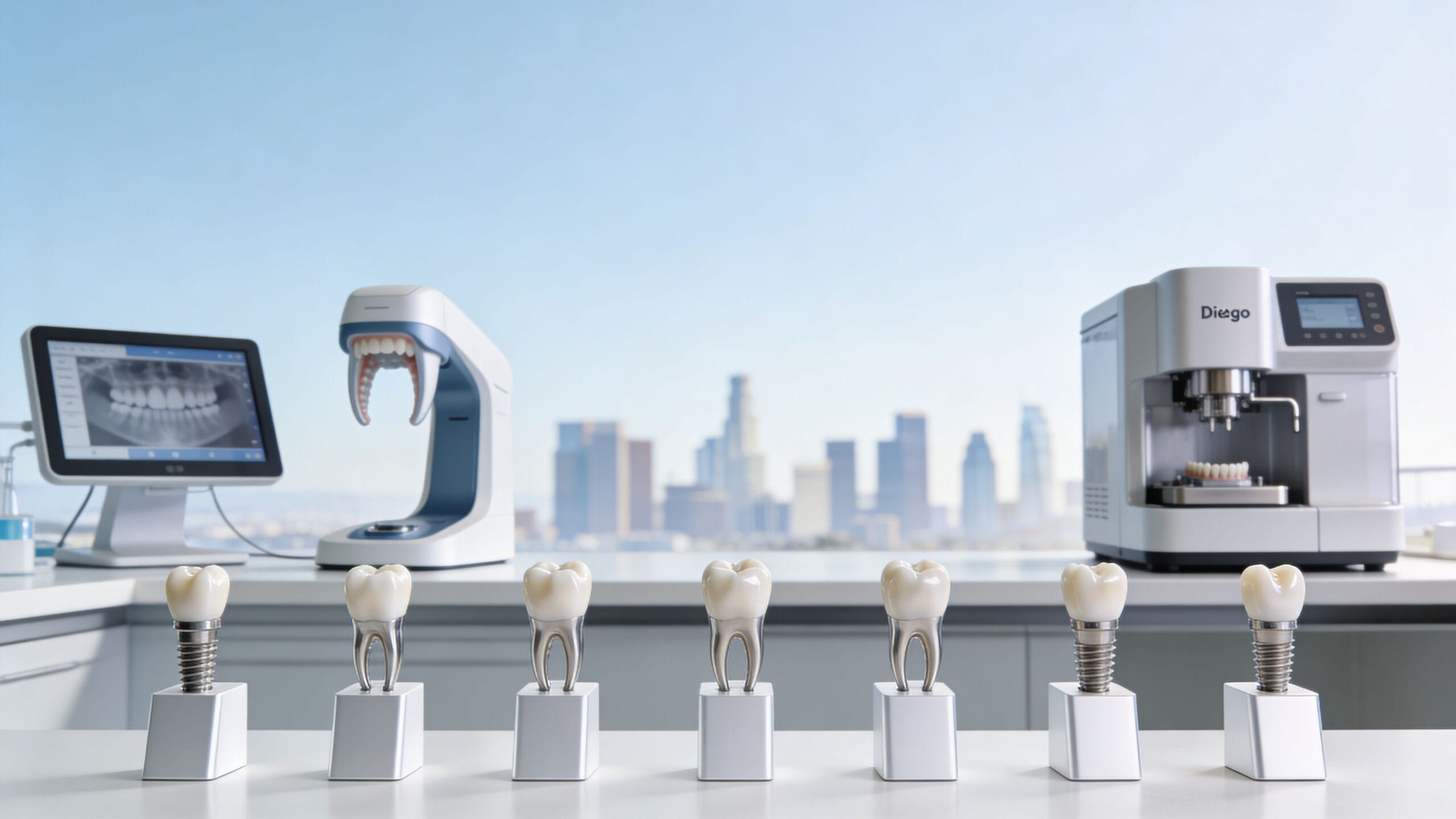
Tooth #7, known as the maxillary right lateral incisor, is the second tooth from the midline in the upper right quadrant of the mouth. It plays a pivotal role in dental aesthetics, speech production, and the shearing of food. Understanding its unique anatomy and common developmental variations is essential for maintaining a functional and confident smile.
In the Universal Numbering System—the standard system used by dental professionals in the United States—each tooth is assigned a specific number from 1 to 32. Tooth #7 is located between the maxillary right central incisor (Tooth #8) and the maxillary right canine (Tooth #6). Because of its prominent position in the "aesthetic zone," it is one of the most frequent subjects of cosmetic dentistry and orthodontic interventions.
The Universal Numbering System and Tooth Identification
To understand a "7 dental overview," one must first grasp how dentists identify teeth. While patients often refer to "the tooth next to the front tooth," clinicians require a standardized nomenclature to ensure accuracy in charting, insurance claims, and surgical procedures.
How Tooth #7 is Located
The Universal Numbering System begins at the upper right third molar (the wisdom tooth), which is designated as #1. The count continues across the upper arch to the upper left third molar (#16). It then drops down to the lower left third molar (#17) and continues around to the lower right third molar (#32). Following this path, Tooth #7 is the second tooth to the right of the midline (from the patient's perspective).
In the International FDI (Fédération Dentaire Internationale) system, which is used globally outside the U.S., Tooth #7 is referred to as Tooth 12. The first digit represents the quadrant (1 for upper right), and the second digit represents the position from the midline (2 for lateral incisor). Despite these different naming conventions, the anatomical structure and clinical importance of the tooth remain identical.
Developmental Timeline
As of 2026, pediatric dental guidelines continue to emphasize the importance of monitoring the eruption of the permanent lateral incisors. Tooth #7 typically erupts between the ages of 8 and 9. It replaces the primary (baby) maxillary right lateral incisor, known as Tooth D in the primary numbering system. Delays in eruption or the premature loss of the primary predecessor can lead to crowding or impaction, necessitating early orthodontic evaluation.
Anatomical Characteristics of the Maxillary Right Lateral Incisor
Tooth #7 is structurally similar to the central incisor but is smaller in all dimensions except for root length, which is often proportionally longer. Its unique morphology contributes to the "smile arc," the curved line created by the edges of the upper teeth.
Crown Morphology
The crown of Tooth #7 is characterized by several distinct surfaces:
- Labial Surface: The front surface facing the lips. It is more rounded than the central incisor, often possessing a convex shape that catches light and defines the tooth's appearance.
- Lingual Surface: The back surface facing the tongue. It features a prominent cingulum (a rounded elevation near the gum line) and a lingual fossa (a shallow depression).
- Incisal Edge: The biting edge. Unlike the flat edge of a central incisor, the lateral incisor often has more rounded "incisal angles," particularly the distal angle (the corner furthest from the midline).
Root Structure
The root of Tooth #7 is typically single and conical. It is often longer than the crown, providing significant stability within the alveolar bone. A common anatomical variation is a slight distal curvature at the apex (the tip of the root), which dentists must account for during root canal therapy or extractions.
Internal Anatomy
Internally, the tooth houses the dental pulp, containing nerves and blood vessels. The pulp chamber of Tooth #7 is generally narrower than that of the central incisor. Because the tooth is relatively thin, it is particularly susceptible to pulp damage from deep decay or physical trauma, which can happen frequently due to its position at the front of the mouth.
Common Clinical Conditions Affecting Tooth #7
Because of its complex developmental origin, Tooth #7 is prone to several unique conditions that can affect both health and appearance.
Peg Laterals (Microdontia)
One of the most common anomalies associated with Tooth #7 is the "peg lateral." This occurs when the tooth develops with a significantly smaller, cone-like shape rather than a full rectangular crown. This is a form of localized microdontia. Peg laterals often result in significant spacing (diastema) between the teeth, which many patients choose to correct for aesthetic reasons.
Congenitally Missing Lateral Incisors
The maxillary lateral incisors (both #7 and #10) are among the most frequently congenitally missing teeth in the human dentition, a condition known as hypodontia. When Tooth #7 fails to develop, the canine (Tooth #6) may drift into the space, or a large gap may remain. Treatment usually involves either orthodontic closure of the space or "space maintenance" to allow for a future dental implant.
Dens Invaginatus (Dens in Dente)
Tooth #7 is the most common tooth to exhibit dens invaginatus, a developmental malformation where the outer surface of the tooth folds inward into the pulp chamber. This creates a deep "pit" that is highly susceptible to bacterial colonization and rapid decay. Early detection through radiographic imaging is vital to prevent premature tooth loss.
Restorative and Cosmetic Solutions
Given its role in the "aesthetic zone," treatments for Tooth #7 focus on balancing function with natural-looking results. In 2026, advancements in porcelain technology and digital smile design have made these restorations more durable and lifelike than ever.
Dental Bonding
For minor chips or small peg laterals, composite bonding is an efficient solution. A tooth-colored resin is applied directly to the enamel and sculpted to the desired shape.
- Pros: Cost-effective, minimally invasive, and completed in one visit.
- Cons: Prone to staining over time and less durable than porcelain.
Porcelain Veneers
Veneers are thin shells of ceramic bonded to the front of the tooth. They are the gold standard for correcting the shape, color, and alignment of Tooth #7. Veneers are particularly effective for masking peg laterals or severe discoloration that does not respond to whitening.
Dental Implants
In cases where Tooth #7 is congenitally missing or lost due to trauma, a dental implant is often the preferred long-term solution. A titanium post is surgically placed in the jawbone to act as a root, followed by the attachment of a custom porcelain crown. This prevents the bone loss that occurs when a tooth is missing and avoids the need to grind down adjacent healthy teeth for a bridge.
Comparison of Treatment Options for Tooth #7
| Feature | Composite Bonding | Porcelain Veneer | Dental Implant |
|---|---|---|---|
| Durability | 3–7 years | 10–15+ years | 25+ years (Lifetime) |
| Invasiveness | Minimal (No drilling) | Moderate (Thin enamel removal) | High (Surgical) |
| Aesthetics | Good (May stain) | Excellent (Stain resistant) | Excellent |
| Cost (Estimated 2026) | $350–$800 | $1,200–$2,500 | $3,500–$6,000 |
| Primary Use | Minor chips/gaps | Reshaping/Peg laterals | Missing teeth |
Orthodontic Considerations and the "Golden Proportion"
Orthodontists pay close attention to Tooth #7 when planning a smile. The concept of the Golden Proportion suggests that the perceived width of the lateral incisor should be approximately 62% of the width of the central incisor when viewed from the front.
If Tooth #7 is too narrow or malpositioned, it can throw off the entire facial symmetry. Orthodontic treatment (such as braces or clear aligners) is often used to ensure there is exactly enough room for a veneer or implant to be placed on Tooth #7 without it looking "crowded" or "too wide."
Furthermore, Tooth #7 is a common site for "ectopic eruption," where the tooth emerges in the wrong position, often behind the lower teeth (a crossbite). Correcting this early is critical to prevent abnormal wear on the incisal edge.
Maintenance and Long-Term Care
Protecting Tooth #7 requires a combination of diligent home care and professional oversight. Because of its location, it is highly visible but also susceptible to specific types of damage.
- Trauma Prevention: Since Tooth #7 is at the front of the mouth, it is frequently injured during sports or falls. Individuals active in contact sports should always wear a professionally fitted mouthguard.
- Hygiene in the Aesthetic Zone: The lingual pit (the small depression on the back of the tooth) can harbor plaque that is difficult to reach with a standard toothbrush. Flossing and the use of an electric toothbrush are essential to prevent "interproximal" cavities between Tooth #7 and its neighbors.
- Nightguards: Patients who grind their teeth (bruxism) often see the first signs of wear on the incisal edges of the lateral incisors. A nightguard can preserve the length and shape of the tooth.
For more information on tooth numbering and standard dental care, the American Dental Association (ADA) provides comprehensive resources on tooth identification and health.
Frequently Asked Questions
Is Tooth #7 a "front tooth"?
Yes, Tooth #7 is considered one of the six "anterior teeth" in the upper arch. Along with the central incisors and canines, it forms the front-facing part of the smile. While not as large as the central incisors, its presence is crucial for a natural and symmetric appearance.
Why is my Tooth #7 smaller than the one on the other side?
This is a common occurrence known as bilateral asymmetry. While many people have "peg laterals" on both sides, it is possible for only one lateral incisor (either #7 or #10) to be undersized. This can be corrected using cosmetic dentistry techniques like bonding or veneers to match the size of the opposite tooth.
What should I do if my Tooth #7 is missing?
If you are missing Tooth #7, you should consult with both an orthodontist and a restorative dentist. The two main options are to close the gap by moving the canine into the #7 position (canine substitution) or to open the gap and place a dental implant or bridge. Each option has different impacts on your bite and long-term facial profile.
Can a root canal be performed on Tooth #7?
Absolutely. Tooth #7 is a frequent candidate for root canal therapy, often due to trauma-induced nerve death or deep decay in the lingual pit. Because the tooth is thin, dentists often recommend a crown or internal bleaching after a root canal to prevent the tooth from becoming brittle or turning dark.
How much does it cost to fix a peg lateral in 2026?
In 2026, the cost depends on the method of restoration. Simple composite bonding for a peg lateral typically ranges from $400 to $900 per tooth. A more permanent porcelain veneer usually costs between $1,500 and $2,800. Prices vary significantly based on geographic location and the expertise of the cosmetic dentist.
Practical Recommendation
If you are concerned about the appearance or health of Tooth #7, the first step is a comprehensive clinical examination including digital radiographs. Early identification of developmental issues like dens invaginatus or microdontia can save thousands of dollars in future restorative costs. For those looking to enhance their smile, consult a dentist who utilizes Digital Smile Design (DSD) to visualize how changes to Tooth #7 will affect your overall facial aesthetics before beginning any treatment.