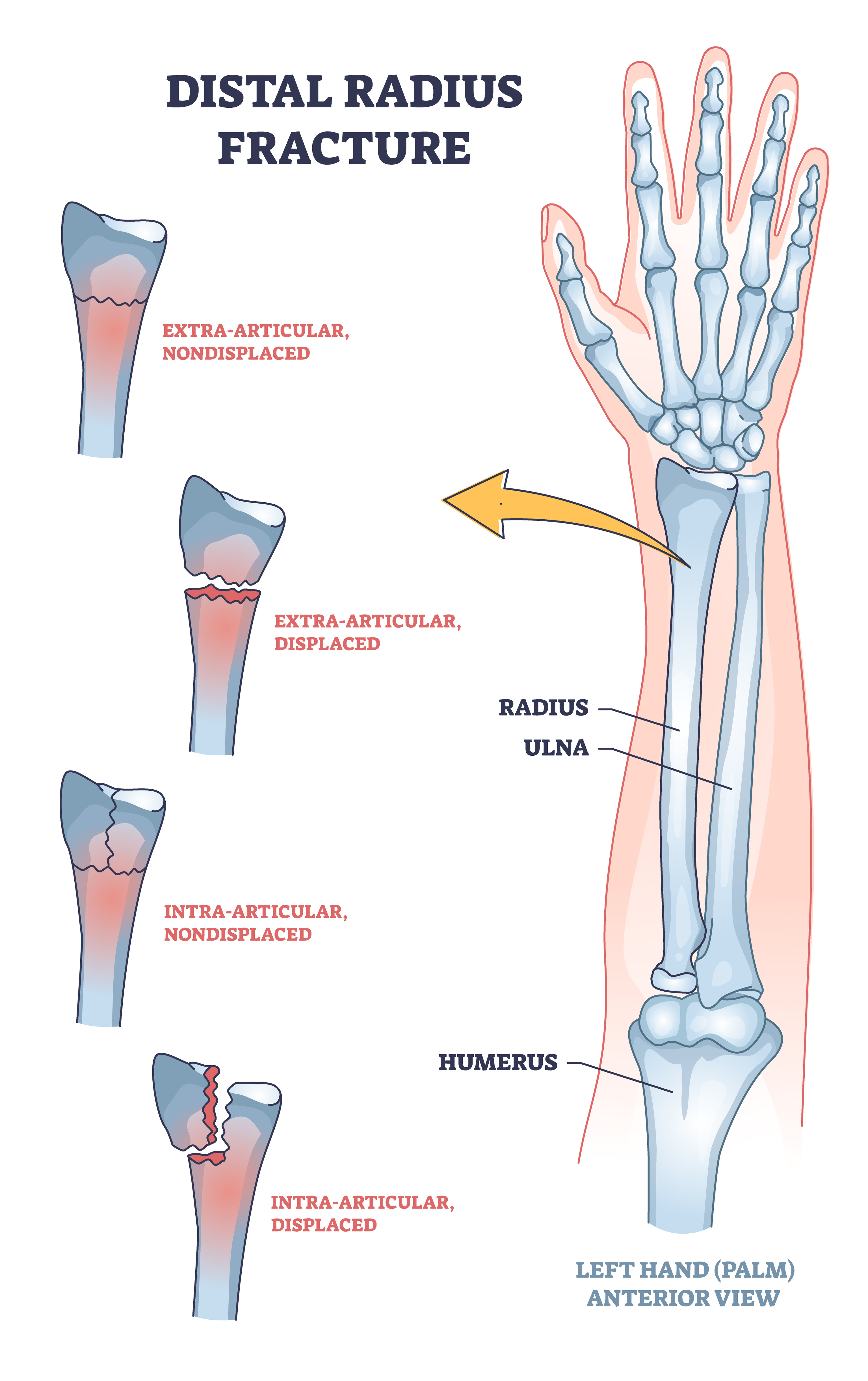
A closed fracture of the distal end of the left radius is a bone break near the wrist on the left arm where the skin remains unbroken. This medical diagnosis involves the larger forearm bone close to the hand, categorized as a closed injury because it does not pierce the cutaneous tissue, significantly reducing immediate infection risks.
Understanding the Anatomy and Medical Terminology
To fully comprehend what a closed fracture of the distal end of the left radius means, it is essential to break down the clinical terminology piece by piece. The radius is one of the two long bones in the forearm, situated on the thumb side, while the ulna occupies the pinky side. The term "distal end" refers to the segment of the bone located farthest from the center of the body or the point of attachment—in this case, the portion of the radius that flares out to form the wrist joint and articulates with the carpal bones of the hand.
A "closed" fracture means that the overlying skin and soft tissues remain intact. Unlike an open or compound fracture, where bone fragments break through the skin surface, a closed injury keeps the internal environment shielded from external pathogens, fundamentally altering the initial management and treatment trajectory. Finally, specifying the "left" radius identifies the precise anatomical location affected, which is vital for surgical planning, documentation, and patient safety protocols.
This specific anatomical region bears significant mechanical load during everyday activities, such as catching oneself during a fall, lifting objects, or twisting a door handle. Because the distal radius contributes significantly to forearm rotation and wrist stability, even a simple fracture in this zone requires careful evaluation to ensure long-term joint function and prevent chronic pain or stiffness.
Common Causes and Mechanics of Injury
The mechanism behind a closed fracture of the distal end of the left radius typically involves high-energy or low-energy trauma applied to an outstretched hand. Healthcare providers frequently refer to this specific injury pattern as a Fall Onto an Outstretched Hand, commonly abbreviated as a FOOSH injury.
When an individual loses balance, the natural defensive reflex is to extend the arms to break the fall. Upon impact, the kinetic energy travels from the palm through the carpal bones and directly into the articular surface and metaphysis of the distal radius. Depending on the exact angle of the wrist at the moment of impact and the bone mineral density of the individual, the resulting break can take various geometric forms, such as extra-articular or intra-articular fractures, transverse breaks, or comminuted fractures where the bone shatters into multiple pieces.
While falls from standing height are the leading cause among older adults—particularly postmenopausal women affected by osteoporosis—high-energy trauma accounts for a significant share of cases in younger demographics. Motor vehicle collisions, bicycle accidents, and sports-related impacts can impart enough force to shatter the distal radius even in patients with optimal bone density. Understanding the specific mechanism helps orthopedic specialists anticipate soft tissue damage, ligamentous injury, and the potential for displacement.
Clinical Presentation and Diagnostic Evaluation
Diagnosing a closed fracture of the distal end of the left radius relies on a combination of physical examination findings and advanced imaging modalities. Patients typically present with immediate, sharp pain at the wrist, localized swelling, tenderness to the touch, and a noticeable decrease in grip strength and range of motion. Depending on whether the bone fragments have shifted, there may also be visible deformity, such as the classic "dinner fork" appearance associated with certain dorsal displacement patterns.
| Diagnostic Step | Purpose and Clinical Significance |
|---|---|
| Physical Examination | Assess neurovascular status, swelling, and deformity; check median nerve function. |
| Standard Radiographs (X-rays) | Confirm the fracture line, displacement, angulation, and articular involvement. |
| Computed Tomography (CT) Scan | Provide detailed 3D views for complex intra-articular fractures requiring surgical planning. |
| Magnetic Resonance Imaging (MRI) | Evaluate associated soft tissue, ligament, and triangular fibrocartilage complex (TFCC) damage. |
During the physical assessment, the examining clinician checks the neurovascular status of the hand. This involves evaluating capillary refill, radial pulse strength, and sensory function in the fingers—particularly testing the median nerve distribution, which runs close to the carpal tunnel and can be compressed by acute swelling or displaced bone fragments. Standard anteroposterior and lateral X-rays are then ordered to visualize the fracture pattern, measure radial tilt, and determine joint congruity. For complex breaks involving the joint surface, a CT scan may be necessary to map out precise surgical reduction strategies.
Classification Systems and Fracture Patterns
Medical professionals utilize specific classification systems to categorize distal radius fractures, guiding treatment decisions and prognosis. Not all breaks at the end of the radius are identical; their stability and orientation dictate whether conservative management will succeed or if surgical intervention is mandatory.
- Colles' Fracture: The most common pattern, characterized by dorsal (backward) displacement of the distal bone fragment, typically resulting from a fall on an outstretched palm.
- Smith's Fracture: Often called a reverse Colles' fracture, featuring volar (forward) displacement of the fragment, usually caused by falling backward onto a flexed wrist.
- Barton's Fracture: An intra-articular fracture accompanied by dislocation or subluxation of the radiocarpal joint, involving either the dorsal or volar rim of the radius.
- Chopart and Die-Punch Fractures: Specialized intra-articular shear injuries that disrupt specific facets of the articular surface where the carpal bones articulate with the radius.
Recognizing these patterns helps clinicians predict the likelihood of post-traumatic arthritis and joint instability. Intra-articular fractures—those extending into the wrist joint—demand higher precision in anatomical reduction to prevent uneven wear on the cartilage, whereas extra-articular fractures may tolerate minor angular discrepancies without long-term functional deficits.
Non-Surgical Treatment Options
When a closed fracture of the distal end of the left radius is undisplaced or stable, non-surgical management represents the primary treatment pathway. The overarching goal of conservative care is to hold the bone fragments in proper alignment while natural osteoblastic activity bridges the fracture gap with new bone.
The standard immobilization method involves applying a well-molded fiberglass or plaster cast that extends from below the elbow to the metacarpal heads, often incorporating a period of initial splinting to accommodate acute swelling. If the fracture is slightly displaced upon presentation, an orthopedic specialist may perform a closed reduction. This procedure involves administering a local anesthetic block or procedural sedation, manipulating the bone back into acceptable alignment manually, and immediately securing the limb in a cast.
Throughout the immobilization period—which typically lasts between six to eight weeks—regular follow-up appointments and serial X-rays are scheduled. These check-ups ensure that the bone does not slip out of alignment inside the cast as initial swelling subsides. Patients are advised to perform gentle active exercises for the fingers, elbow, and shoulder to prevent stiffness in adjacent joints while the wrist remains locked.
Surgical Intervention and Fixation Techniques
Surgical management becomes necessary when a closed fracture of the distal end of the left radius is unstable, significantly displaced, severely comminuted, or involves a disrupted articular surface that cannot be satisfactorily restored or maintained via closed manipulation.
Several surgical techniques exist, tailored to the specific fracture geometry and patient characteristics:
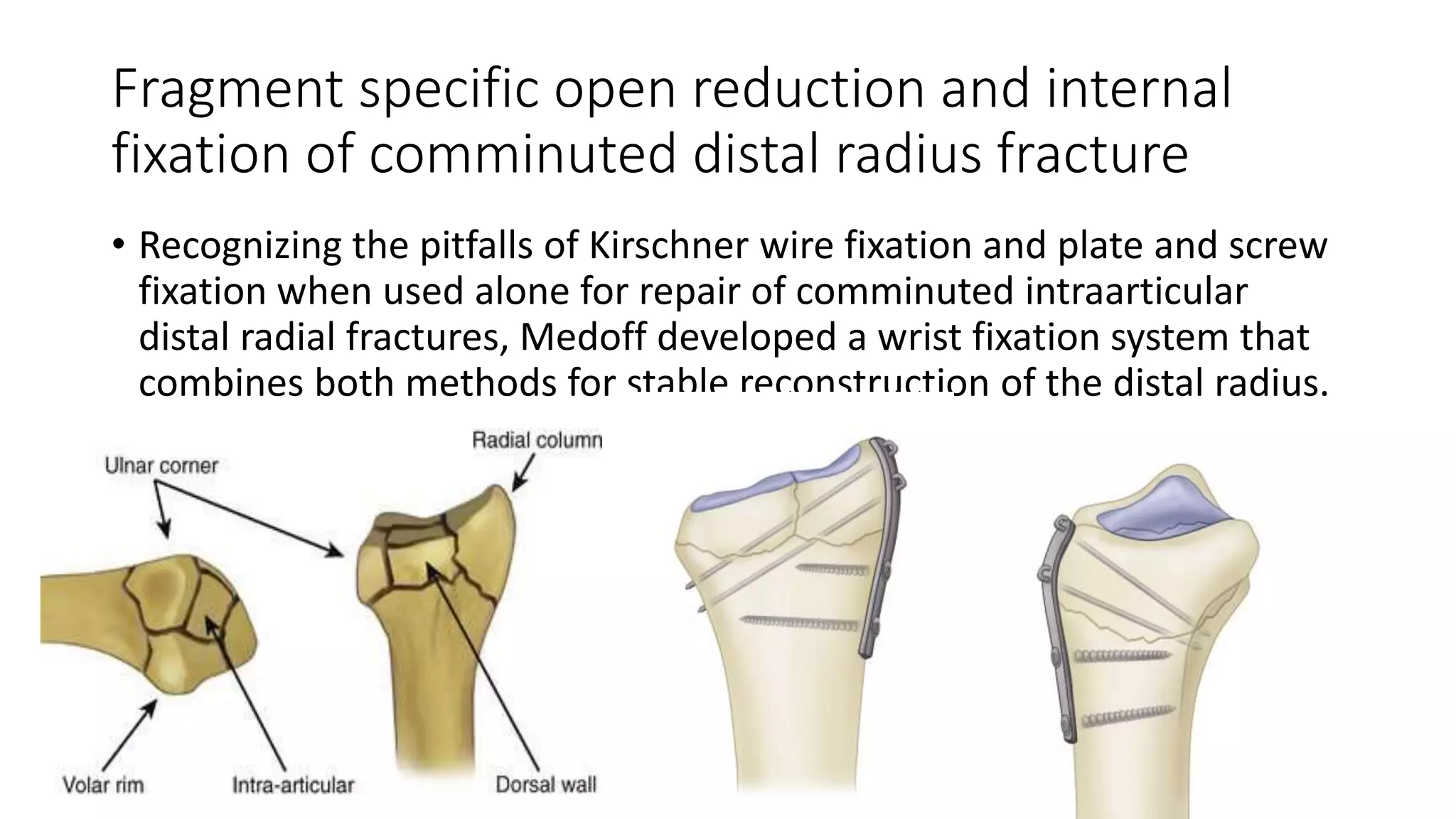
- Closed Reduction and Percutaneous Pinning (CRPP): The surgeon manipulates the bone fragments externally and inserts metal pins (Kirschner wires) through the skin across the fracture site to hold it stable beneath a protective cast.
- Open Reduction and Internal Fixation (ORIF): The surgeon makes an incision—typically on the volar (palmar) side of the wrist—to directly visualize the bone fragments, align them anatomically, and secure them using a low-profile titanium plate and locking screws.
- External Fixation: A frame placed outside the wrist with pins inserted into the radius above the break and into the metacarpal bones below, utilizing ligamentotaxis (tension on surrounding soft tissues) to pull the fracture fragments into place.
Internal fixation with a volar locking plate has become a gold standard for many displaced distal radius fractures because it provides rigid stability, allowing patients to begin early wrist mobilization therapy shortly after the operation, thereby minimizing long-term joint stiffness.
Rehabilitation, Physical Therapy, and Recovery
Recovery from a closed fracture of the distal end of the left radius extends well beyond the removal of a cast or the healing of the bone itself. Soft tissue contractures, muscle atrophy, and joint stiffness invariably develop during the immobilization phase, making structured physical therapy an indispensable component of successful rehabilitation.
Once the treating physician confirms adequate bone healing—verified through clinical examination and follow-up imaging—rehabilitation focuses on restoring active and passive range of motion. Therapists utilize gentle stretching exercises, scar tissue mobilization, and joint mobilizations to loosen the radiocarpal and midcarpal joints. As flexibility improves, progressive strengthening programs are introduced, incorporating resistance bands, grip-strengthening devices, and weight-bearing exercises to rebuild forearm musculature.
Full recovery is a gradual process. While most patients resume basic activities of daily living within three months of injury or surgery, achieving maximum grip strength, endurance, and weather-resistant comfort can take up to a year. Adherence to home exercise programs and gradual progression of load-bearing activities are critical to preventing re-injury and optimizing functional outcomes.
Potential Complications and Long-Term Outlook
Although the vast majority of patients heal successfully from a closed fracture of the distal end of the left radius, several potential complications warrant monitoring by both the patient and the clinical team.
- Malunion or Nonunion: Malunion occurs when the bone heals in an abnormal position, potentially leading to altered wrist mechanics, chronic pain, or cosmetic deformity. Nonunion, though rare in the distal radius due to its rich blood supply, happens when the bone fails to unite entirely.
- Post-Traumatic Arthritis: Damage to the articular cartilage of the radiocarpal joint during the initial impact can accelerate cartilage degradation over time, resulting in degenerative joint disease.
- Complex Regional Pain Syndrome (CRPS): A chronic, progressive nerve condition characterized by severe, disproportionate burning pain, swelling, and vasomotor changes in the hand and wrist.
- Nerve Compression: Acute swelling or surgical scarring can compress the median nerve inside the carpal tunnel, causing carpal tunnel syndrome symptoms such as numbness, tingling, and weakness in the thumb, index, and middle fingers.
With modern diagnostic tools, advanced surgical hardware, and targeted rehabilitation protocols, the long-term prognosis for this injury is generally favorable. Most individuals regain a high functional capacity, allowing them to return to work, sports, and daily routines with minimal residual limitation.
Frequently Asked Questions
How long does a closed fracture of the distal end of the left radius take to heal?
The bone typically heals in approximately six to eight weeks of immobilization or surgical fixation. However, complete functional recovery, including the restoration of full strength, flexibility, and endurance, can take anywhere from six months to a full year depending on age, overall health, and adherence to physical therapy.
Is surgery always required for this type of wrist fracture?
No, surgery is not always necessary. Undisplaced or stable fractures are successfully managed non-surgically using closed reduction and cast immobilization. Surgery is reserved for unstable, displaced, or intra-articular breaks that require mechanical stabilization to ensure proper joint alignment and function.
Can I move my fingers while wearing a cast for a left distal radius fracture?
Yes, moving your fingers, thumb, and elbow is strongly encouraged while in a cast or splint. Regular movement prevents joint stiffness, reduces swelling, and maintains circulation in the hand and forearm during the healing phase.
What is the difference between a closed fracture and an open fracture?
In a closed fracture, the skin and soft tissues overlying the broken bone remain intact, shielding the internal injury from external contamination. In an open fracture, the bone breaks through the skin, creating a direct pathway for bacteria and elevating the risk of deep bone infection.
When should I contact a doctor during my recovery?
You should contact your orthopedic specialist or seek immediate medical attention if you experience severe, unmanaged pain, unrelenting numbness or tingling in your fingers, a bluish or pale discoloration of the fingertips, or if your cast becomes excessively tight, cracked, or loose.
Next Actions and Medical Disclaimer
If you or someone you know has sustained a suspected wrist injury, the immediate priority is to seek professional medical evaluation through urgent care, an emergency department, or an orthopedic specialist. Obtain diagnostic imaging promptly to confirm or rule out a fracture and prevent improper healing.
Disclaimer: This article is for informational and educational purposes only and does not constitute formal medical advice, diagnosis, or treatment. Always consult a qualified healthcare provider regarding any musculoskeletal injury or medical condition.