CD4 flow cytometry is the gold-standard diagnostic laboratory method used to quantify CD4+ T-lymphocytes in peripheral blood. Primarily utilized for staging HIV infection and monitoring the efficacy of antiretroviral therapy (ART), this technique employs laser-based technology and fluorescently labeled antibodies to identify and count specific immune cells, guiding critical clinical decisions regarding immune health and opportunistic infection prophylaxis.
Understanding the Biological Role of CD4+ T-Lymphocytes
To appreciate the necessity of CD4 flow cytometry, one must first understand the role of the target cell. CD4+ T-lymphocytes, often called "helper" T cells, are the orchestrators of the human immune response. These cells do not directly kill pathogens; instead, they recognize foreign antigens presented by Major Histocompatibility Complex (MHC) class II molecules on the surface of antigen-presenting cells.
Once activated, CD4 cells secrete cytokines that signal other immune components—such as CD8+ cytotoxic T cells, B-lymphocytes, and macrophages—to respond to the threat. In the context of Human Immunodeficiency Virus (HIV), the virus specifically targets and destroys these CD4+ cells. As the CD4 count drops, the immune system loses its ability to coordinate a defense, leading to Acquired Immunodeficiency Syndrome (AIDS) and making the body susceptible to opportunistic infections and certain cancers.
By 2026, while viral load testing has become the primary measure of treatment success, the CD4 count remains the essential baseline indicator of an individual's immunological status and their immediate risk of clinical complications.
The Mechanics of Flow Cytometry in Immunology
Flow cytometry is a sophisticated analytical technique that measures the physical and chemical characteristics of particles (usually cells) as they flow in a fluid stream through a beam of light. For a CD4 overview, the process relies on three integrated systems: fluidics, optics, and electronics.
The Fluidics System
The fluidics system's purpose is to transport cells in a single-file line to the "interrogation point," where the laser beam hits them. This is achieved through a process called hydrodynamic focusing. A sample of the patient's blood is injected into a faster-moving "sheath fluid." The pressure differential forces the cells into a narrow central core, ensuring that only one cell passes through the laser at a time. This precision is vital for an accurate "absolute count," which is the number of CD4 cells per microliter (mm³) of blood.
The Optics System
The optics system consists of lasers to illuminate the cells and optical filters to direct the resulting light signals to the appropriate detectors. When a laser hits a cell, the light is scattered in two main ways:
- Forward Scatter (FSC): Correlates with the cell's size.
- Side Scatter (SSC): Correlates with the cell's internal complexity or granularity.
In CD4 flow cytometry, however, the most important data comes from fluorescence. Before the blood is run through the machine, it is incubated with monoclonal antibodies conjugated to fluorescent dyes (fluorochromes). These antibodies are designed to bind specifically to the CD4 molecule on the surface of T cells. When the laser excites these dyes, they emit light at specific wavelengths, which the optics system captures.
The Electronics System
The electronics system converts the light signals into electronic signals that a computer can process. The intensity of the fluorescence is proportional to the number of CD4 molecules on the cell surface, allowing the software to distinguish CD4+ T cells from other cell types, such as monocytes (which express CD4 at lower levels) or CD8+ T cells.
Clinical Indications for CD4 Flow Cytometry
While HIV management is the most common use case, CD4 flow cytometry is utilized in several clinical scenarios in 2026:
- HIV Staging and Baseline Assessment: Every patient newly diagnosed with HIV receives a CD4 count to determine the severity of immune suppression. This helps clinicians decide if the patient needs immediate prophylaxis for infections like Pneumocystis pneumonia (PCP) or Cryptococcosis.
- Monitoring Antiretroviral Therapy (ART): For patients on ART, an increasing CD4 count indicates "immunologic reconstitution." If the count fails to rise despite an undetectable viral load, it may suggest "discordant response," requiring closer clinical monitoring.
- Primary Immunodeficiencies: Infants or adults suspected of having genetic immune defects (such as SCID) require flow cytometry to characterize lymphocyte subsets.
- Organ Transplantation: Patients receiving immunosuppressive therapy to prevent organ rejection undergo CD4 monitoring to ensure their immune system isn't suppressed to a point of extreme vulnerability.
- Chemotherapy Monitoring: Certain intensive chemotherapy regimens significantly deplete T cell populations; CD4 counts help determine when it is safe to stop prophylactic antibiotics.
The Laboratory Procedure: From Phlebotomy to Data Analysis
A standard CD4 flow cytometry overview must include the practical steps taken in a clinical pathology lab to ensure results are accurate and reproducible.
1. Sample Collection and Preparation
Blood is typically collected in an EDTA (lavender top) tube. It is critical that the sample is not refrigerated, as cold temperatures can alter the cell surface markers and affect the "gating" of the lymphocytes. For absolute counts, the sample must ideally be processed within 24 to 48 hours.
2. Immunophenotyping (Staining)
A specific volume of whole blood is mixed with a "cocktail" of fluorochrome-labeled antibodies. A common panel is the "4-color" or "6-color" panel, which includes:
- CD45: To identify all white blood cells (leukocytes).
- CD3: To identify all T-lymphocytes.
- CD4: To identify helper T cells.
- CD8: To identify cytotoxic T cells.
3. Erythrocyte Lysis
Since red blood cells (erythrocytes) outnumber white blood cells by about 1,000 to 1, they would "swamp" the detectors. A lysing solution is added to the sample to dissolve the red blood cells while leaving the white blood cells intact for analysis.
4. Gating and Analysis
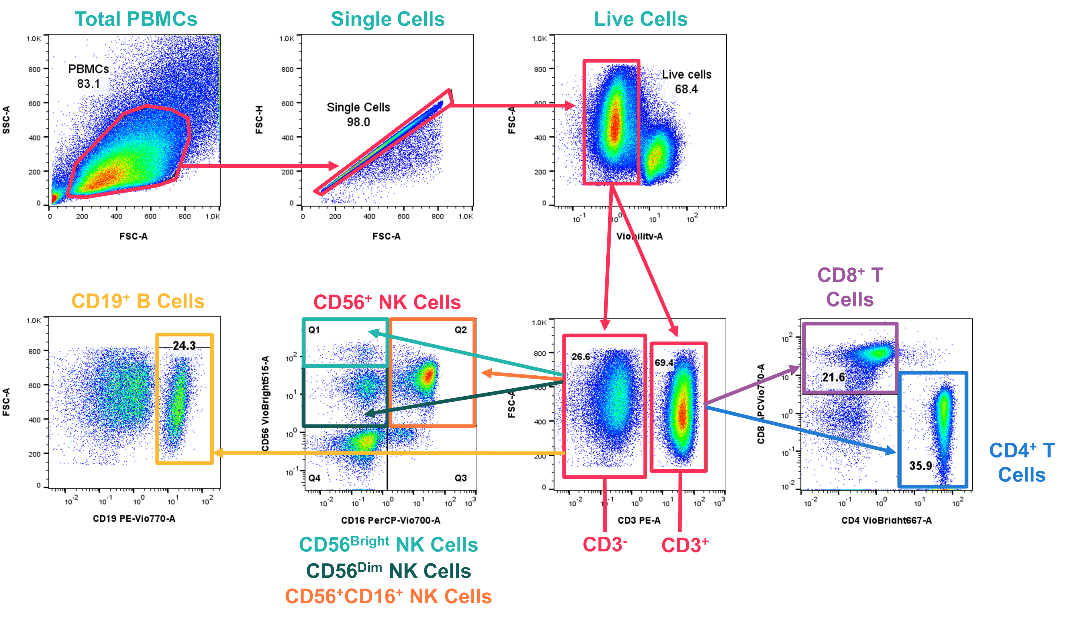
Laboratory technicians use software to perform "gating." This is the process of drawing boundaries around specific populations of cells on a scatter plot.
- First, the software identifies lymphocytes based on their size (FSC) and low granularity (SSC).
- Next, it isolates CD3+ cells (T cells) from that group.
- Finally, it identifies the percentage of those T cells that are CD4+.
Interpreting CD4 Results: Reference Ranges and Thresholds
The interpretation of CD4 flow cytometry involves two primary metrics: the Absolute CD4 Count and the CD4 Percentage.
The Absolute Count is calculated by multiplying the total White Blood Cell (WBC) count by the percentage of lymphocytes, then by the percentage of CD4+ T cells. In 2026, many labs use "single-platform" technology where counting beads are added to the sample, allowing the machine to calculate the absolute count directly without needing an external hematology analyzer.
| CD4 Count (cells/mm³) | Immune Status Category | Clinical Significance / Action Required |
|---|---|---|
| 500 – 1,500 | Normal / Strong | Normal immune function in healthy adults. |
| 350 – 499 | Mild Suppression | Generally asymptomatic; monitoring for ART response. |
| 200 – 349 | Moderate Suppression | Increased risk of common infections; ART highly recommended. |
| < 200 | Severe Suppression (AIDS) | High risk for opportunistic infections (PCP, Toxoplasmosis). |
| < 50 | Advanced Disease | Extreme risk for CMV and Mycobacterium avium complex (MAC). |
Medical Disclaimer: These ranges are general guidelines. Clinical decisions must be made by a qualified healthcare provider based on the individual patient's history and local 2026 health protocols.
The Importance of the CD4 Percentage
In some cases, the absolute count can fluctuate due to factors like hydration, stress, or minor infections. The CD4 percentage (the percentage of total lymphocytes that are CD4+) is often more stable. A CD4 percentage below 14% is clinically equivalent to an absolute count below 200 cells/mm³, even if the absolute number appears higher due to an elevated total WBC count.
Technical Variations: Single-Platform vs. Dual-Platform
There are two primary ways laboratories arrive at the final CD4 number. Understanding these is essential for interpreting longitudinal data, especially if a patient switches laboratories.
- Dual-Platform Method: This was the traditional standard. It requires two different machines: a flow cytometer to get the CD4 percentage and a hematology analyzer to get the total lymphocyte count. The absolute CD4 count is then calculated manually or by software. This method has a higher margin of error because it combines the variability of two different instruments.
- Single-Platform Method: This is the preferred standard in 2026. This method uses a single flow cytometer that either uses a known volume of blood or internal "counting beads" (fluorescent microspheres). Because the count is derived from one instrument, the precision is much higher, and the results are more consistent across different testing dates.
Factors Influencing CD4 Count Accuracy
CD4 counts are dynamic. A single "low" reading does not always indicate a clinical decline. Several factors can cause transient changes in results:
- Diurnal Variation: CD4 counts are typically lowest in the morning and peak in the evening. For consistency, patients should ideally have their blood drawn at the same time of day for every follow-up.
- Acute Non-HIV Infections: A common cold, the flu, or a flare-up of another condition can temporarily depress the CD4 count.
- Physical Stress and Exercise: Intense physical exertion shortly before a blood draw can cause a temporary spike or drop in circulating lymphocytes.
- Medications: Certain drugs, such as corticosteroids or chemotherapy agents, directly affect lymphocyte levels independently of HIV progression.
- Laboratory Variability: Even with automation, there is an inherent "coefficient of variation" (CV) in flow cytometry, meaning a change of 10-15% between tests might just be statistical noise rather than a biological change.
2026 Advances in Point-of-Care (POC) CD4 Testing
In 2026, the landscape of flow cytometry has expanded from centralized "high-throughput" laboratories to decentralized Point-of-Care (POC) settings. These are smaller, often cartridge-based devices that can be used in rural clinics or resource-limited environments.
While traditional flow cytometers require specialized technicians and stable electricity, POC CD4 devices use simplified fluidics and fixed optical paths. These devices provide results in under 30 minutes, allowing for "same-day" clinical decisions. This is particularly vital for the "Rapid ART Initiation" protocols used in 2026, where patients start treatment immediately upon diagnosis.
Frequently Asked Questions
1. How often should a CD4 count be performed?
In 2026, for a patient with a stable, undetectable viral load and a CD4 count consistently above 500 cells/mm³ for over a year, testing may only be required annually or may even be suspended at the physician's discretion. However, for those starting ART or those with low counts, testing is typically done every 3 to 6 months until the immune system stabilizes.
2. Can a CD4 count increase after it has dropped?
Yes. With effective Antiretroviral Therapy (ART) that suppresses the HIV viral load, the body can produce new CD4+ T cells, and the "memory" T cells can redistribute. This process is called immune reconstitution. While the count may not always return to "normal" (above 500), most patients see a significant increase that removes them from the risk zone for opportunistic infections.
3. What is the difference between CD4 and Viral Load?
The CD4 count measures the damage already done to the immune system (the "defense"), while the viral load measures the activity of the virus (the "attacker"). The goal of treatment is to lower the viral load to "undetectable" levels, which then allows the CD4 count to rise or stay stable.
4. Why is my CD4 percentage normal but my absolute count low?
This often happens if your total white blood cell count is low (leukopenia). If you have fewer white blood cells overall, the absolute number of CD4 cells will be lower even if the "mix" or percentage of those cells is healthy. In these cases, doctors usually look at the CD4 percentage as a more reliable indicator of immune health.
5. Does a low CD4 count always mean I have AIDS?
Technically, according to the CDC, an HIV-positive individual is diagnosed with AIDS if their CD4 count drops below 200 cells/mm³ or if they develop a specific "AIDS-defining" opportunistic infection. However, with 2026 medical treatments, the "AIDS" label is less of a permanent status and more of a clinical stage that can be managed and reversed with proper medication.
Summary and Next Actions
CD4 flow cytometry remains an indispensable pillar of modern immunology and HIV care. By providing a precise snapshot of the body's cellular defenses, it enables healthcare providers to tailor treatments, prevent life-threatening infections, and monitor the long-term health of patients with immune-mediated conditions.
Recommended Next Actions:
- For Patients: If your CD4 count is currently below 200 cells/mm³, ensure you are strictly adherent to your ART regimen and discuss opportunistic infection prophylaxis (such as Bactrim or Septra) with your provider. Always try to schedule your blood draws for the same time of day.
- For Clinicians: When interpreting a sudden drop in CD4 count, always check the patient’s viral load and screen for recent acute illnesses or high-stress events before assuming treatment failure. Rely on the CD4 percentage if the absolute count seems inconsistent with the patient's clinical presentation.