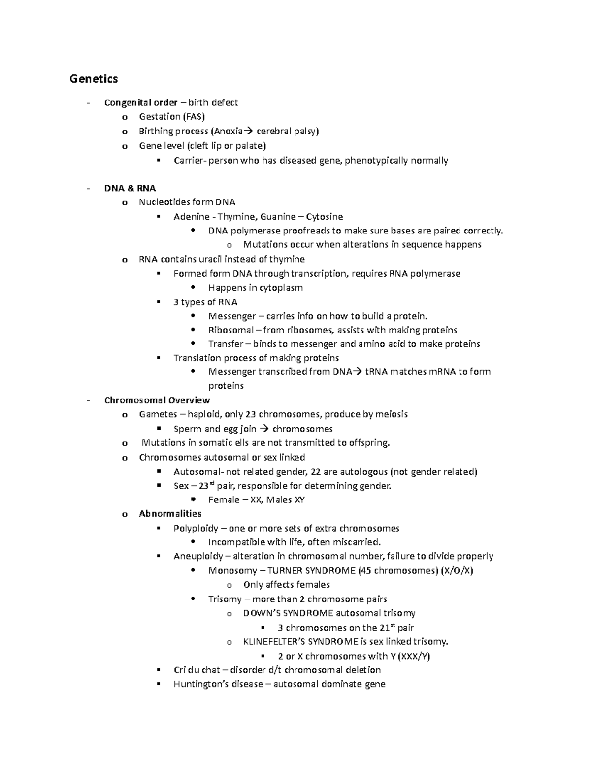
Anoxia at birth occurs when a newborn baby experiences a total deprivation of oxygen to the brain and bodily tissues during the labor, delivery, or immediate post-birth period. This medical emergency interrupts vital oxygen flow, triggering rapid cellular damage and potential brain injury known as hypoxic-ischemic encephalopathy (HIE), requiring immediate, specialized medical resuscitation and interventions like therapeutic hypothermia.
Medical Disclaimer: This article is intended solely for educational and informational purposes and does not constitute medical advice, diagnosis, or treatment. Medical decisions regarding neonatal care, diagnosis, and post-discharge treatment must always be made in consultation with qualified healthcare professionals, neonatologists, and pediatric specialists.
Defining Anoxia at Birth: Anoxia vs. Hypoxia and HIE
To understand the impact of oxygen deprivation during delivery, it is essential to distinguish between related medical terms:
- Hypoxia: A partial reduction in the oxygen supply reaching tissues and organs. While serious, some tissue function may be maintained temporarily.
- Anoxia: The complete absence or total deprivation of oxygen reaching the body’s cells and organs. Anoxia represents the most severe end of the oxygen-deprivation spectrum.
- Asphyxia: A physiological condition characterized by simultaneous oxygen deprivation (hypoxia/anoxia) and excessive carbon dioxide accumulation (hypercapnia) in the blood, leading to severe metabolic acidosis.
- Hypoxic-Ischemic Encephalopathy (HIE): The specific neurological brain dysfunction caused by severe hypoxia or anoxia combined with reduced blood flow (ischemia) to the brain.
When a fetus or newborn experiences anoxia, the brain is deprived of metabolic fuel. The primary phase of injury occurs immediately as cellular energy production fails, leading to an influx of sodium and calcium into brain cells, cellular swelling, and cell death.
If circulation is restored, a secondary phase of injury—known as reperfusion injury—often unfolds over the following 6 to 48 hours. During this phase, inflammatory reactions, toxic chemical releases (such as excess glutamate), and cellular apoptosis (programmed cell death) can cause further damage to previously surviving neural tissue.
Key Causes and Risk Factors During Delivery
Anoxia at birth rarely occurs without underlying intrapartum or antepartum triggers. Oxygen is transferred from the mother’s bloodstream through the placenta and umbilical cord directly to the fetus. Any interruption along this metabolic pipeline can induce severe oxygen failure.
Umbilical Cord Complications
- Umbilical Cord Prolapse: Occurs when the cord slips into the birth canal ahead of the baby. As the baby descends during contractions, the cord is compressed between the baby's body and the birth canal, blocking blood flow.
- Severe Nuchal Cord: When the umbilical cord wraps tightly around the infant's neck multiple times or forms a true knot, restricting arterial and venous blood delivery.
- Cord Compression: Mechanical squeezing of the cord due to low amniotic fluid (oligohydramnios) or physical impingement.
Placental Complications
- Placental Abruption: The premature separation of the placenta from the uterine wall before delivery, rapidly cutting off oxygen and blood flow to the fetus.
- Placenta Previa: A condition where the placenta partially or completely covers the cervix, risking catastrophic maternal and fetal hemorrhage during labor.
- Placental Insufficiency: Chronic degradation of placental blood vessels, often secondary to maternal preeclampsia or chronic hypertension, which leaves the infant with poor metabolic reserve to tolerate normal labor contractions.
Maternal and Uterine Factors
- Uterine Rupture: A catastrophic tear of the uterine wall—most frequently occurring along a prior Cesarean section scar—causing massive internal maternal blood loss and immediate interruption of fetal oxygenation.
- Maternal Shock or Hypotension: Extreme drops in maternal blood pressure caused by systemic infection (sepsis), heavy bleeding, or epidural complications.
- Intra-amniotic Infection (Chorioamnionitis): Bacterial infection of the fetal membranes and amniotic fluid, causing systemic inflammation that exacerbates fetal brain vulnerability to oxygen deprivation.
Delivery Complications
- Prolonged or Arrested Labor: Extended pressure on the infant’s head and prolonged uterine contractions reduce inter-contraction recovery time for placental blood replenishment.
- Shoulder Dystocia: An emergency where the baby's head emerges but the shoulders become impacted behind the mother's pubic bone, delaying delivery and causing severe umbilical cord compression.
Recognizing the Symptoms of Birth Anoxia
Healthcare teams monitor both fetal heart rate traces during labor and the physical condition of the baby immediately upon delivery to identify signs of oxygen deprivation.
| Immediate at Delivery | Early Neurological (First 24 Hrs) |
|---|---|
| • Apgar score 0–3 past 5 minutes | • Neonatal seizures |
| • Cyanosis (blue/pale skin tone) | • Lethargy, stupor, or coma |
| • Weak or completely absent gasping | • Hyperalertness or irritability |
| • Severe hypotonia ("floppy baby") | • Weak or absent suck/swallow |
| • Weak or absent heart rate | • Abnormal posture / rigid tone |
Physical Signs at Delivery
- Low Apgar Scores: An Apgar score evaluated at 1, 5, and 10 minutes assesses heart rate, respiratory effort, muscle tone, reflex irritability, and skin color. Scores below 3 at 5 and 10 minutes strongly suggest severe asphyxia.
- Depressed Respiration: The infant may be apneic (not breathing) or display weak, irregular gasping efforts.
- Cyanosis or Pallor: Skin tone appears pale, blue, or gray due to severe lack of oxygenated blood circulating through peripheral tissues.
- Severe Hypotonia: The baby appears abnormally limber or "floppy," exhibiting little to no spontaneous movement or resistance to passive movement.
Early Neurological Symptoms
In the hours following delivery, an infant affected by anoxia may exhibit neurological signs indicative of brain injury:
- Neonatal Seizures: Abnormal brain electrical activity manifesting as repetitive twitching, cycling movements of the limbs, subtle eye-rolling, or prolonged apnea episodes.
- Altered Consciousness: Ranging from hyperalertness and extreme irritability (in mild cases) to profound lethargy, stupor, or coma (in severe cases).
- Reflex Abnormalities: Absent or blunted Moro (startle) reflex, sucking reflex, and swallowing reflex.
Emergency Diagnostics and Clinical Evaluation
Accurate diagnosis of anoxia at birth requires a combination of laboratory testing, physical neurological examinations, and advanced neuroimaging.
Umbilical Cord Blood Gas Analysis
Immediately following delivery of an affected baby, blood is drawn from the umbilical artery to measure blood gases. Key markers indicating severe birth asphyxia include:
- Arterial pH: A blood pH lower than 7.00 indicates severe acidemia.
- Base Deficit: A base deficit of 12 mmol/L or higher indicates significant metabolic acidosis caused by anaerobic metabolism in oxygen-starved tissues.
Sarnat Staging System
Pediatricians and neonatologists utilize the Sarnat Staging System to categorize the severity of neonatal encephalopathy into mild (Stage 1), moderate (Stage 2), or severe (Stage 3). This clinical assessment evaluates alertness, muscle tone, autonomic reflexes, and seizure activity to help guide treatment decisions.
Neuroimaging Modalities
- Cranial Ultrasound: Often performed bedside in the Neonatal Intensive Care Unit (NICU) during the first hours of life to rule out structural malformations or major intracranial hemorrhages.
- Magnetic Resonance Imaging (MRI): The gold-standard diagnostic tool for evaluating neonatal brain injury. Brain MRIs are typically performed between day 4 and day 14 of life (after completing therapeutic cooling). Diffusion-Weighted Imaging (DWI) can identify specific patterns of brain injury, such as damage to the basal ganglia, thalamus, or watershed cortex regions.
Electroencephalography (EEG and aEEG)
Continuous amplitude-integrated EEG (aEEG) or full multichannel EEG monitoring is applied in the NICU to detect subclinical seizure activity (seizures without physical twitching) and evaluate background brain wave patterns. Severe background abnormalities, such as burst suppression or flat traces, correlate with more significant brain injury.
Immediate Treatment and Therapeutic Interventions
Management of birth anoxia relies on emergency resuscitation at delivery followed by specialized neuroprotective strategies in a tertiary NICU.
- Anoxia / Birth Asphyxia
- Immediate Resuscitation (NRP Protocols)
- Airway clearance, ventilation, oxygenation
- Identify Candidates for Therapeutic Hypothermia
- Age ≥35-36 weeks, ≤6 hours from delivery, metabolic acidosis & encephalopathy
- Whole-Body or Head Cooling for 72 Hours
- Target core temperature: 33.5°C (92.3°F)
- Controlled Rewarming Phase (12 Hours)
- Slow rewarming at 0.5°C per hour to 36.5°C
- Post-Cooling Assessment & Rehabilitation
- Brain MRI, EEG, neurodevelopmental follow-up
- Immediate Resuscitation (NRP Protocols)
Neonatal Resuscitation Program (NRP)
The immediate management of an anoxic baby follows standard NRP guidelines:
- Airway Management: Clearing fluid or meconium from the airway if obstructing respiration.
- Positive Pressure Ventilation (PPV): Administering artificial breaths using a bag-valve-mask or endotracheal tube to establish functional residual capacity in the lungs.
- Circulatory Support: Performing chest compressions and administering intravenous epinephrine if the infant’s heart rate remains below 60 beats per minute despite effective ventilation.
Therapeutic Hypothermia (Cooling Therapy)
Therapeutic hypothermia is the standard of care for infants born at or near term (typically $\ge 35$ weeks gestation) who present with moderate-to-severe HIE. According to clinical consensus guidelines published by the American Academy of Pediatrics (AAP), cooling therapy must be initiated within 6 hours of birth to be effective.
- Mechanism: Lowering core body temperature to 33.5°C (92.3°F) slows cellular metabolism, reduces free radical formation, inhibits programmed cell death (apoptosis), and decreases cerebral inflammation.
- Duration: The baby is maintained on a cooling blanket for exactly 72 hours, followed by a gradual rewarming process over 12 hours (at a rate of approximately 0.5°C per hour).
- Clinical Impact: Clinical trials have demonstrated that therapeutic hypothermia significantly reduces the rate of mortality and severe neurodevelopmental disability at 18 to 24 months of age.
Neurocritical Care Support
Alongside cooling therapy, the NICU care team provides comprehensive supportive medical care:
- Seizure Control: Anti-seizure medications such as phenobarbital, levetiracetam, or fosphenytoin are administered intravenously to stop seizure activity and prevent secondary brain damage.
- Hemodynamic Stabilization: Inotropic support (e.g., dopamine, dobutamine) is used to maintain adequate blood pressure and cerebral perfusion pressure.
- Respiratory Management: Mechanical ventilation ensures optimal oxygen saturation while avoiding hyperoxia (too much oxygen) or hypocapnia (excessively low carbon dioxide), both of which can worsen brain tissue injuries.
Long-Term Effects and Complications
The long-term prognosis for a baby who survives anoxia at birth depends on the severity of oxygen deprivation, the promptness of medical intervention, and the specific regions of the brain affected.
Cerebral Palsy (CP)
Anoxia is a recognized cause of dyskinetic, spastic, or ataxic cerebral palsy. Damage to the basal ganglia, motor cortex, or corticospinal tracts disrupts the brain's ability to control muscle movement, tone, and posture.
Cognitive and Developmental Delays
Children affected by neonatal anoxia may experience varying degrees of intellectual disability, learning disabilities, executive dysfunction, and memory impairments as they reach school age.
Epilepsy and Seizure Disorders
Surviving neurons in injured regions of the brain may form abnormal electrical connections, resulting in chronic epilepsy that requires long-term anticonvulsant medication management.
Sensory Deficits
- Cortical Visual Impairment (CVI): Visual dysfunction caused by damage to the visual processing centers in the brain's occipital lobes rather than an issue with the eyes themselves.
- Sensorineural Hearing Loss: Damage to the auditory pathways or auditory cortex resulting from hypoxia and ischemic injury.
Behavioral and Psychological Outcomes
Children with a history of birth anoxia face higher rates of Attention-Deficit/Hyperactivity Disorder (ADHD), sensory processing disorders, and social-emotional challenges.
Diagnostic and Clinical Comparison Table
The following table summarizes the diagnostic features, neurological staging criteria, and clinical trajectories of infants presenting with neonatal encephalopathy following an anoxic event.
| Clinical Parameter | Stage 1: Mild Encephalopathy | Stage 2: Moderate Encephalopathy | Stage 3: Severe Encephalopathy |
|---|---|---|---|
| Level of Consciousness | Hyperalert, irritable | Lethargic or obtunded | Stuporous, comatose |
| Muscle Tone | Normal or slightly increased | Hypotonia (floppy) | Severe flaccidity |
| Suck / Swallow Reflex | Weak or uncoordinated | Absent or severely depressed | Absent |
| Seizure Activity | None | Common (focal or multifocal) | Severe, frequent, or status epilepticus |
| Umbilical Arterial pH | 7.00 – 7.15 | 6.85 – 6.99 | < 6.85 |
| Base Deficit | 10 – 15 mmol/L | 16 – 20 mmol/L | > 20 mmol/L |
| aEEG / EEG Pattern | Normal or continuous low voltage | Discontinuous or discontinuous pattern | Burst suppression or flat/isoelectric |
| Cooling Therapy Indication | Typically not indicated | Indicated (Initiate < 6 hours) | Indicated (Initiate < 6 hours) |
| Typical Long-Term Outcome | Good; high likelihood of full recovery | Variable; 30–50% risk of neurodisability | High risk of mortality or severe CP/disability |
Therapies, Early Intervention, and Family Resources
When an infant experiences birth anoxia, structured early intervention programs can optimize long-term neurodevelopmental outcomes by capitalizing on early neuroplasticity.
Early Intervention Programs (EIP)
In the United States, state-funded Early Intervention programs (authorized under Part C of the Individuals with Disabilities Education Act) offer specialized developmental support for infants from birth to age three. Services are provided in the home or community settings.
Rehabilitation Therapies
- Physical Therapy (PT): Focuses on gross motor skills, joint mobility, muscle tone management, and head control.
- Occupational Therapy (OT): Helps children master fine motor skills, feeding, sensory integration, and self-care routines.
- Speech-Language Pathology (SLP): Addresses oral-motor weakness, swallowing safety (dysphagia management), and early communication skill development.
Clinical Follow-Up Care
Children surviving birth anoxia require ongoing care coordination through specialized pediatric high-risk infant follow-up clinics, involving pediatric neurologists, developmental-behavioral pediatricians, physiatrists, and orthopedists.
For research and clinical updates on brain injury care, parents can refer to resources provided by the National Institute of Neurological Disorders and Stroke (NINDS).
Frequently Asked Questions
What is the difference between anoxia and hypoxia at birth?
Hypoxia refers to a partial reduction in oxygen delivery to bodily tissues, whereas anoxia represents a total lack of oxygen. Anoxia is the most severe stage of oxygen deprivation and rapidly leads to cellular death if blood flow and oxygen supply are not immediately restored.
How quickly does brain damage occur during anoxia at birth?
Irreversible brain cell injury can begin within 4 to 6 minutes of complete oxygen deprivation. However, the extent of damage depends on whether blood supply (ischemia) is completely stopped, the baby's metabolic reserve, and how quickly effective medical resuscitation measures are initiated.
What is therapeutic hypothermia, and how does it help?
Therapeutic hypothermia, or cooling therapy, involves safely lowering a newborn’s core body temperature to 33.5°C (92.3°F) for 72 hours. Initiated within 6 hours of birth, it slows brain cell metabolism, limits inflammation, and reduces the risk of death or long-term neurodevelopmental disability from HIE.
Can a baby fully recover from anoxia at birth?
Yes, full recovery is possible, particularly in cases of mild-to-moderate anoxia where effective resuscitation and cooling therapy are delivered promptly. However, infants with severe, prolonged anoxia face higher risks of long-term challenges such as cerebral palsy, cognitive delays, or epilepsy.
What diagnostic tests confirm if anoxia occurred during delivery?
Doctors use umbilical cord blood gas analysis (checking for pH < 7.00 and high base deficit), bedside EEG/aEEG continuous brain wave monitoring, and brain MRI scans (typically conducted 4 to 14 days after birth) to confirm the diagnosis and assess the extent of brain tissue injury.
Recommended Next Steps for Affected Families
If your baby has experienced anoxia at birth, navigating medical care can feel overwhelming. Consider taking the following practical steps:
- Request Full Medical Records: Obtain complete copies of maternal admission charts, fetal heart rate monitoring strips, labor logs, delivery notes, umbilical cord gas analyses, and NICU discharge summaries.
- Ensure Referral to a High-Risk Infant Follow-Up Clinic: Schedule regular multi-disciplinary evaluations with pediatric neurologists and developmental specialists to monitor motor, cognitive, and sensory milestones.
- Enroll in Early Intervention Services: Contact your state or local Early Intervention agency immediately after hospital discharge to set up home visits for physical, occupational, and speech therapy.
- Connect with Family Support Networks: Reach out to established non-profit organizations such as Hope for HIE or United Cerebral Palsy for peer support, educational materials, and guidance.
- Seek Independent Medical Evaluation: If you have questions regarding the management of your labor, fetal distress monitoring, or emergency delivery response times, consider requesting a clinical review by an independent specialist.