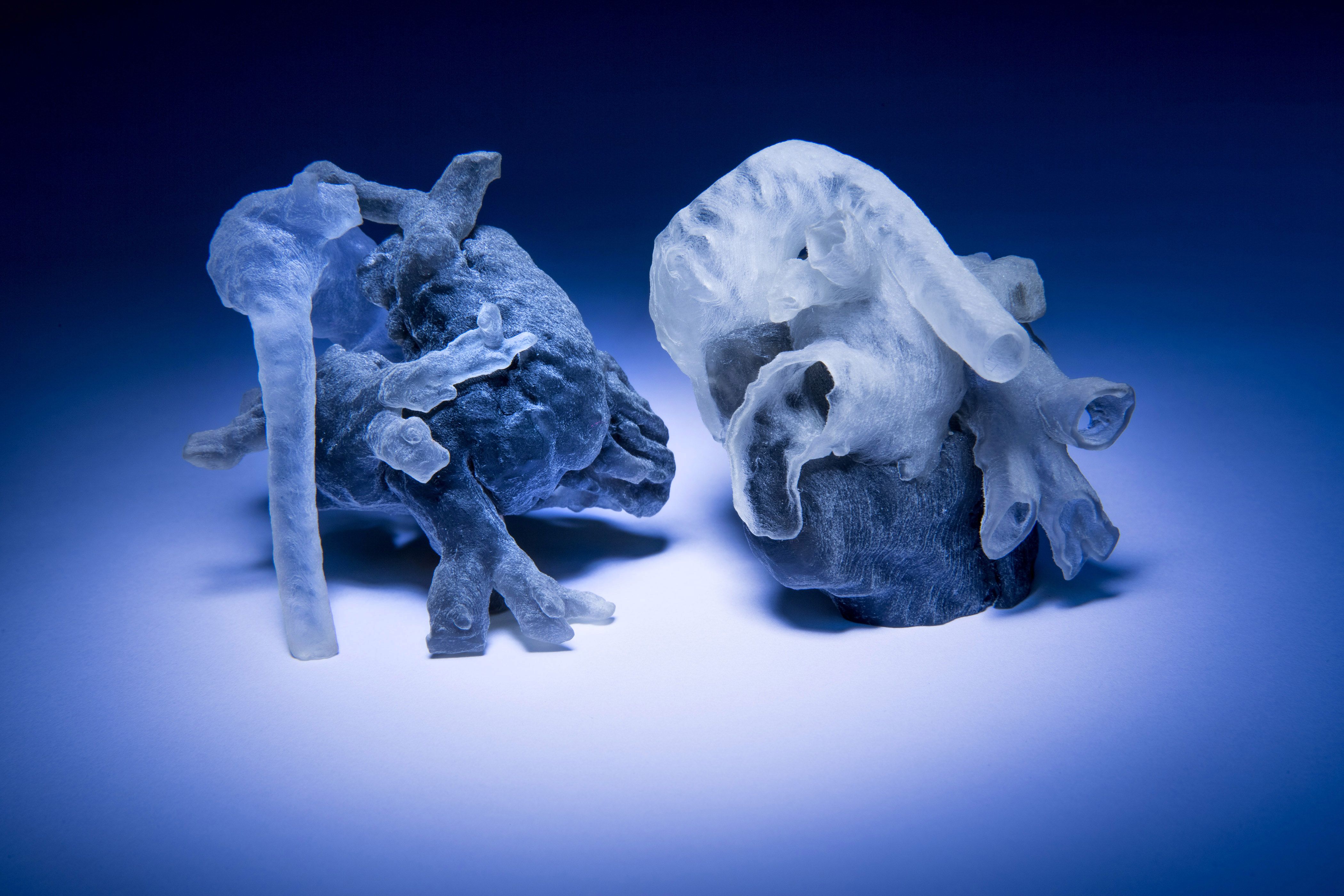
Utilizing 3D printing heart models for surgery allows cardiothoracic surgeons and congenital heart specialists to translate complex medical imaging data into tangible, patient-specific anatomical replicas, facilitating precise surgical planning, procedural rehearsal, and intraoperative orientation for complex cardiovascular interventions.
Introduction and Clinical Context
Cardiovascular surgery demands absolute precision. Historically, surgeons relied on two-dimensional computed tomography (CT) scans, magnetic resonance imaging (MRI), and echocardiograms, mentally reconstructing complex three-dimensional anatomies before entering the operating room. While effective for many routine procedures, congenital heart defects, intricate valvular pathologies, and complex aneurysms often present geometric challenges that defy mental 3D visualization.
The advent of medical-grade additive manufacturing—commonly known as 3D printing—has revolutionized this paradigm. By converting standard Digital Imaging and Communications in Medicine (DICOM) files derived from patient scans into computer-aided design (CAD) models, clinical teams can print exact replicas of a patient’s heart. These models allow medical professionals to touch, manipulate, and literally dissect the pathology before making a single incision. This guide explores the complete workflow, technical requirements, materials, clinical applications, and limitations associated with 3D printing heart models for surgery.
Disclaimer: The following information is intended for educational and informational purposes regarding medical technology and clinical workflows. It does not constitute medical, surgical, or professional healthcare advice.
The Clinical Value Proposition: Why Patient-Specific Models Matter
The primary driver behind 3D printing in cardiac surgery is risk reduction through enhanced preparation. Complex structural heart diseases, such as Tetralogy of Fallot, transposition of the great arteries, and complex ventricular septal defects, feature unique spatial relationships that vary significantly from patient to patient.
Improving Preoperative Planning
When a surgeon holds a physical, patient-specific replica of a malformed heart, the benefits are immediate. Surgical strategies can be simulated directly on the model. For instance, in valve-sparing aortic root replacement or complex congenital repairs, the team can measure wall thicknesses, assess the precise distance between critical structures like the conduction system and defect margins, and pre-fit prosthetic devices such as patches, rings, or transcatheter valves.
Intraoperative Guidance and Communication
Beyond the operating theater, these models serve as vital communication tools. Multidisciplinary heart teams—comprising cardiac surgeons, interventional cardiologists, imaging specialists, and anesthesiologists—can review the physical model together to reach a consensus on the surgical approach. Furthermore, these models enhance patient and family education, helping lay individuals understand the nature of the condition and the proposed surgical intervention, thereby alleviating anxiety and improving informed consent.
The End-to-End Workflow: From Scan to Scalpel
Transforming a digital medical scan into a physical, sterilizable heart model requires a rigorous, multi-step pipeline. Each phase demands precision to ensure the final physical model accurately reflects the patient's true anatomy.
- DICOM Imaging (CT/MRI)
- Image Segmentation & 3D Reconstruction
- CAD Optimization & Mesh Repair
- 3D Printing & Fabrication
- Post-Processing, Cleaning, & Sterilization
- Clinical Application in Surgery
1. Medical Imaging Acquisition
The process begins with high-resolution diagnostic imaging. Contrast-enhanced computed tomography (CT) and magnetic resonance imaging (MRI) are the gold standards. The acquisition protocol must be optimized specifically for 3D printing, featuring thin slices (typically less than 1 millimeter) and high signal-to-noise ratios to ensure clear differentiation between blood pools, myocardial tissue, and adjacent structures.
2. Image Segmentation and Reconstruction
Once the DICOM data is acquired, it is imported into specialized medical segmentation software (such as Materialise Mimics, Simpleware, or Horos). Segmentation is the process of isolating the region of interest—in this case, the heart chambers, great vessels, and coronary arteries—from surrounding tissues based on Hounsfield unit thresholds (in CT scans) or signal intensities (in MRIs). The software then aggregates these segmented voxels into a surface mesh, typically exported as an STL (Standard Tessellation Language) file.
3. Computer-Aided Design (CAD) and Optimization
The raw STL file often contains digital artifacts, "noise," or holes that require manual cleaning by a biomedical engineer or clinical specialist. During this phase, the digital model may be hollowed out to save material, split along anatomical planes to allow internal visualization of heart valves and septal defects, or modified to include mounting fixtures.
4. 3D Printing Technologies and Materials
Different surgical applications require different physical properties. A model intended for visual inspection differs significantly from one intended for tactile surgical cutting and suturing.
| Printing Technology | Common Materials | Primary Clinical Use Case | Mechanical Properties |
|---|---|---|---|
| Material Jetting (PolyJet) | Vero, Tango, Agilus30 | Multi-material complex congenital defects, soft-tissue simulation | High dimensional accuracy; capable of blending rigid and elastomer components. |
| Stereolithography (SLA) | Photopolymer resins (Clear, Flexible) | Visual anatomical review, sizing, rigid structural assessment | Smooth surface finish; excellent clarity for internal chamber visualization. |
| Fused Deposition Modeling (FDM) | PLA, ABS, TPU | Educational displays, cost-effective basic anatomical overviews | Economical; lower resolution and visible layer lines compared to PolyJet/SLA. |
| Selective Laser Sintering (SLS) | Polyamide (Nylon) | Robust structural models, surgical guides, heavy handling | High mechanical strength; typically opaque. |
5. Post-Processing and Sterilization
Once printed, models undergo cleaning to remove support materials—such as dissolving wax supports in an ultrasonic bath for PolyJet prints. Depending on institutional protocols and whether the model will enter the sterile field during an operation, the model must undergo appropriate sterilization. Common methods include hydrogen peroxide gas plasma or ethylene oxide (EtO), as many 3D printing polymers cannot withstand the high temperatures of standard steam autoclaves without warping.
Technical Considerations and Limitations
Despite its profound clinical utility, 3D printing heart models for surgery presents several technical, logistical, and financial challenges that healthcare institutions must navigate.
Material Limitations vs. Real Tissue
While advanced elastomers can closely mimic the elastic modulus of relaxed myocardium or vascular walls, no synthetic polymer perfectly replicates the complex, anisotropic, viscoelastic behavior of living, beating, or bleeding human cardiac tissue. Surgeons must account for these material differences when practicing incisions, traction, or suturing on printed models.
Time and Cost Factors
Producing a patient-specific heart model is resource-intensive. The total turnaround time—from image acquisition and segmentation to printing, post-processing, and sterilization—typically ranges from 24 to 72 hours. This limits the utility of 3D printing in acute emergency cardiovascular interventions, reserving it primarily for elective or planned complex procedures. Furthermore, specialized medical-grade 3D printers, software licenses, certified biocompatible materials, and dedicated personnel contribute to significant capital and operational expenses.
Regulatory and Quality Assurance Standards
In many jurisdictions, 3D-printed anatomical models used for direct clinical decision-making are subject to stringent regulatory oversight (such as FDA clearance in the United States or MDR compliance in the European Union). Hospitals establishing in-house 3D printing laboratories must implement robust quality management systems to validate every step of the workflow, ensuring dimensional fidelity and preventing segmentation errors that could misinform a surgical plan.
Frequently Asked Questions
How long does it take to 3D print a patient-specific heart model?
The entire workflow generally takes between 24 and 72 hours. Image segmentation and digital cleanup take a few hours, actual printing can range from 4 to 20 hours depending on the technology and model complexity, and post-processing, washing, curing, and sterilization add additional time.
Are these models printed in actual size (1:1 scale)?
Yes. For surgical planning and device sizing, models are almost universally printed at an exact 1:1 scale to ensure that measurements, valve sizes, and spatial relationships match the patient's true anatomy.
Can 3D printed heart models be taken directly into the operating room?
Yes, provided they have been printed using biocompatible materials and subjected to a validated hospital-approved sterilization protocol (such as hydrogen peroxide gas plasma). Surgeons can reference them on the sterile back table during the procedure.
Do insurance companies cover the cost of medical 3D printing?
Reimbursement policies vary significantly by geographic region, healthcare system, and specific clinical indication. While coverage for structural heart and congenital planning is expanding, institutions often navigate specific billing codes or absorb costs as part of advanced clinical care initiatives.
Can a 3D printed heart model simulate blood flow?
Standard anatomical models are static. However, advanced engineering laboratories can print compliant, hollow models and integrate them into mock circulatory loops using pulsatile pumps and blood-mimicking fluids to test catheter navigation and hemodynamics in vitro.
Practical Recommendations and Next Actions
For clinical teams and healthcare institutions looking to integrate 3D printing into their cardiovascular surgery programs, a phased approach is recommended:
- Establish a Multidisciplinary Team: Combine the expertise of cardiothoracic surgeons, biomedical engineers, radiologists, and perfusionists.
- Start with External Partnerships: Before investing in capital-intensive in-house medical printers, utilize established medical 3D printing service bureaus to outsource model production and validate clinical workflows.
- Implement Standard Operating Procedures (SOPs): Define clear protocols for image acquisition parameters, segmentation software validation, cleaning, and sterilization to maintain patient safety and regulatory compliance.