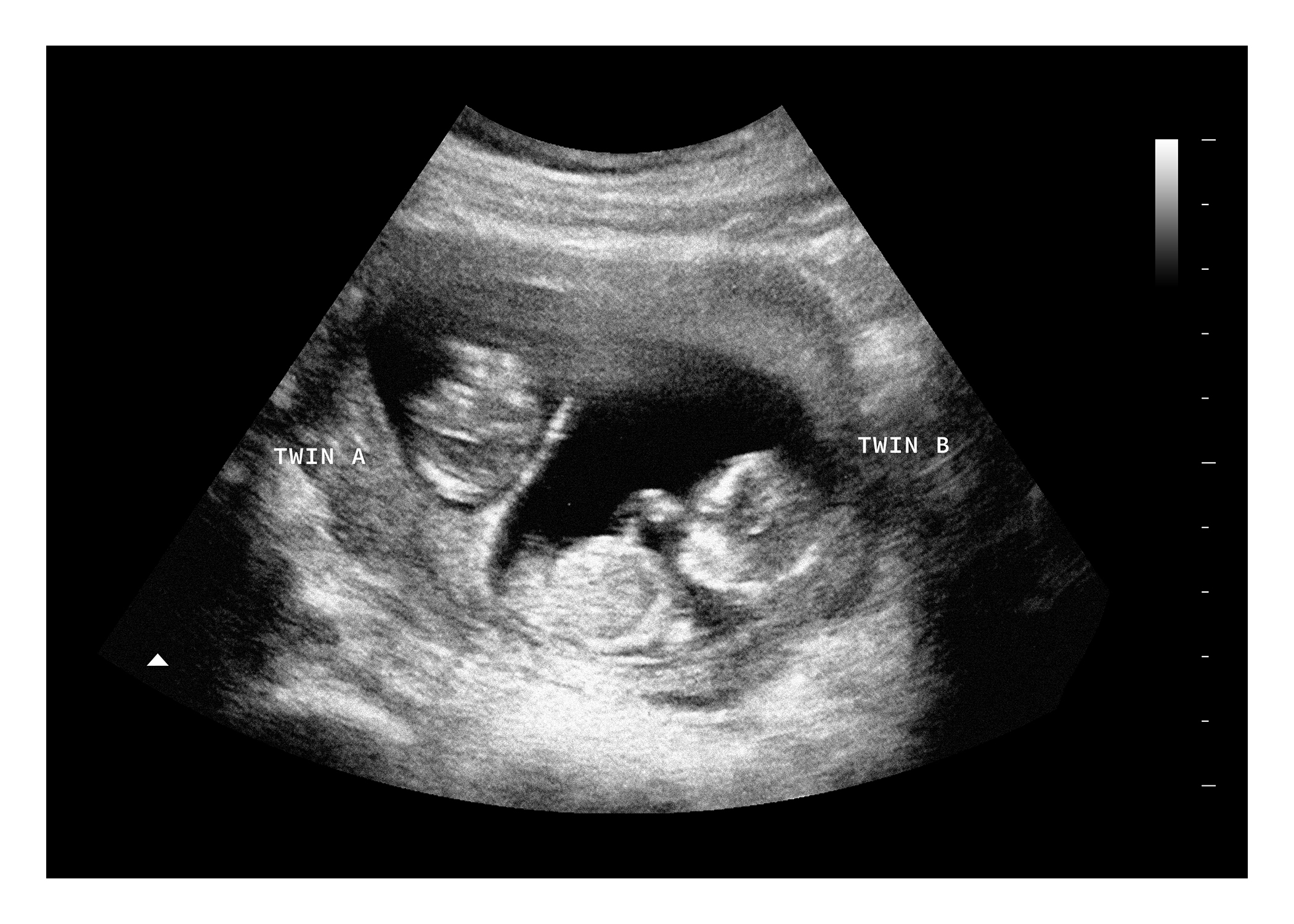
A 10-week ultrasound for identical twins is a critical early milestone used to evaluate structural development, confirm gestational age, and—most importantly—determine chorionicity and amnionicity by mapping the number of placentas and amniotic sacs.
Understanding the Purpose of an Early First-Trimester Scan
The first trimester of a twin pregnancy marks a period of rapid embryonic development and high-stakes clinical assessment. While a routine dating scan can occur anywhere from 8 to 13 weeks of gestation, a targeted ultrasound at precisely 10 weeks offers an optimal window for high-resolution imaging. At this stage, the embryos measure approximately 30 to 40 millimetres from crown to rump, allowing specialized obstetric sonographers and maternal-fetal medicine (MFM) subspecialists to visualize fine anatomical structures that remain obscure during earlier weeks.
For parents expecting identical, or monozygotic, twins, this scan serves a dual purpose. Beyond confirming viability and dating the pregnancy accurately, the clinical team focuses intensely on establishing the placental and sac configuration. Unlike fraternal twins, who always develop separate placentas and sacs, identical twins arise from a single fertilized egg that splits. How and when that split occurs dictates whether the pregnancy is classified as monochorionic-diamniotic (sharing one placenta, separate sacs) or monochorionic-monoamniotic (sharing both a single placenta and a single sac), or in rare cases, conjoined. Identifying these details by week 10 is essential because monochorionic pregnancies carry unique risks that require specialized, frequent surveillance throughout gestation.
Determining Chorionicity and Amnionicity: The "Lambda" and "T" Signs
The single most critical piece of clinical information derived from a 10-week twin ultrasound is the determination of chorionicity. Chorionicity refers to the number of placentas, whereas amnionicity refers to the number of amniotic sacs. In identical twin pregnancies, establishing these factors early dictates the entire management protocol of the prenatal journey.
| Clinical Feature | Monochorionic Twins |
|---|---|
| Placentas | Single Shared Placenta |
| Amniotic Sacs | Two Sacs (Diamniotic) or One (Mono) |
| Dividing Membrane Thickness | Thin (< 2 mm) or Absent |
| Sign at Placental Base | "T-Sign" (Perpendicular junction) |
| Recommended Surveillance Interval | Every 2 Weeks starting at 16 wks |
Sonographers look at the junction where the dividing membrane meets the placenta to categorize the pregnancy. In dichorionic pregnancies, a thick ridge of tissue known as the "lambda sign" (or twin-peak sign) is visible where the two placental masses merge. Conversely, identical twins typically display the "T-sign," where the thin inter-twin membrane inserts into the placenta at a 90-degree angle without projecting extra tissue.
Furthermore, the thickness of the dividing membrane is measured with high-frequency transvaginal probes. A thin membrane—often measuring less than two millimetres—strongly supports a monochorionic-diamniotic diagnosis. If no membrane is visualized at all between the two active embryos, and they share a single fluid space, a monochorionic-monoamniotic (mono-mono) gestation is suspected. Mono-mono pregnancies require intense inpatient or highly vigilant outpatient monitoring due to the risk of umbilical cord entanglement.
Technical Specifications and Imaging Modalities at 10 Weeks
Achieving diagnostic clarity at 10 weeks requires specific technical protocols. Transabdominal ultrasonography provides a broad overview of the pelvic cavity, but transvaginal ultrasonography is universally preferred for detailed first-trimester twin evaluations. Transvaginal probes operate at higher frequencies, bringing the transducer closer to the uterus and yielding superior resolution of embryonic cardiac activity, early limb buds, and the delicate inter-twin membrane.
During the scan, the sonographer captures standardized biometric measurements and structural markers:
- Crown-Rump Length (CRL): Measured for each twin independently to verify that growth is tracking synchronously. A significant discrepancy in CRL at 10 weeks can sometimes serve as an early indicator of uneven placental sharing.
- Nuchal Translucency (NT): While the standard window for NT screening is 11 to 13 weeks and 6 days, a preliminary assessment of fluid accumulation behind the fetal neck can sometimes be visualized at 10 weeks, though official aneuploidy risk calculations typically await the late first-trimester scan.
- Embryonic Heart Rate (FHR): Normal embryonic heart rates at 10 weeks typically range between 120 and 160 beats per minute. Both twins are evaluated separately to document active cardiac function.
- Yolk Sac and Gestational Sac Evaluation: The presence, size, and appearance of the secondary yolk sacs are documented to assess early nutritional transfer and rule out blighted ovum or early embryonic demise of one twin.
Clinical Implications of Monochorionicity
Because nearly all identical twins share a placenta, they are subject to unique vascular communications within the placental mass. Blood vessels from both twins interconnect on the shared placental surface, which can lead to hemodynamic imbalances. Identifying a monochorionic placenta at 10 weeks establishes the foundation for future risk mitigation.
The most notable condition associated with monochorionic identical twins is Twin-to-Twin Transfusion Syndrome (TTTS). In TTTS, blood is transferred disproportionately from one donor twin to the recipient twin through shared placental anastomoses. While TTTS typically manifests in the mid-trimester (most commonly between 16 and 26 weeks), establishing the monochorionic baseline at 10 weeks ensures that the patient is immediately funneled into high-risk obstetric care pathways. Early identification prevents delayed diagnoses and allows maternal-fetal medicine specialists to schedule serial growth and fluid-volume scans starting early in the second trimester.
Patient Preparation and What to Expect During the Procedure
Undergoing a 10-week ultrasound for identical twins involves specific preparation steps to ensure optimal image acquisition. Patients are typically instructed to arrive with a moderately full bladder if a transabdominal scan is performed first, though a transvaginal scan often requires an empty bladder for patient comfort and clearer visualization of deep pelvic structures.
The procedure itself usually lasts between 30 and 45 minutes, slightly longer than a singleton scan because the sonographer must meticulously map out Twin A and Twin B, document their relative positions (anterior vs. posterior, left vs. right), and trace the inter-twin membrane from top to bottom. Sonographers use standardized labeling conventions—such as assigning Twin A to the fetus closest to the internal cervical os—to ensure consistency across subsequent serial scans. Following the examination, a qualified physician or radiologist reviews the images to discuss the confirmed chorionicity, estimated due date, and recommended follow-up schedule with the patient.
Frequently Asked Questions
Can identical twins look like fraternal twins on a 10-week ultrasound?
Yes, in very rare instances, an early scan can be misinterpretingly read if the placentas are fused so closely that they mimic dichorionic twins. However, at 10 weeks, a skilled sonographer utilizing high-resolution transvaginal equipment can almost always differentiate the T-sign of monochorionic twins from the lambda sign of dichorionic twins. If ambiguity remains, follow-up scans at 12 to 14 weeks provide definitive clarification.
Why is determining chorionicity at 10 weeks so important?
Chorionicity dictates the entire medical management plan for a twin pregnancy. Monochorionic identical twins share a placenta and are at risk for complications like Twin-to-Twin Transfusion Syndrome and selective intrauterine growth restriction. Knowing this status at 10 weeks ensures appropriate referral to maternal-fetal medicine specialists and timely initiation of specialized monitoring protocols.
What is the difference between diamniotic and monoamniotic identical twins?
Diamniotic identical twins share a single placenta but have separate amniotic sacs divided by a membrane. Monoamniotic identical twins share both the single placenta and the exact same amniotic sac without any dividing membrane. Monoamniotic pregnancies are much rarer and require intensive inpatient or outpatient surveillance due to the risk of umbilical cord entanglement.
Is a transvaginal ultrasound safe at 10 weeks of pregnancy?
Yes, transvaginal ultrasound is a safe, routine, and standard diagnostic procedure during the first trimester. It utilizes harmless sound waves rather than ionizing radiation and allows clinicians to obtain much clearer, higher-resolution images of the embryos and placental membranes than is possible with abdominal scanning alone at this early stage.
Next Steps and Professional Guidance
Patients who receive a confirmed diagnosis of a monochorionic identical twin pregnancy at their 10-week ultrasound should request a formal consultation with a maternal-fetal medicine (MFM) specialist or high-risk obstetrician. Establishing a coordinated care plan early ensures that subsequent monitoring schedules—typically involving bi-weekly ultrasound surveillance starting around 16 weeks—are properly arranged to safeguard maternal and fetal health throughout the pregnancy.