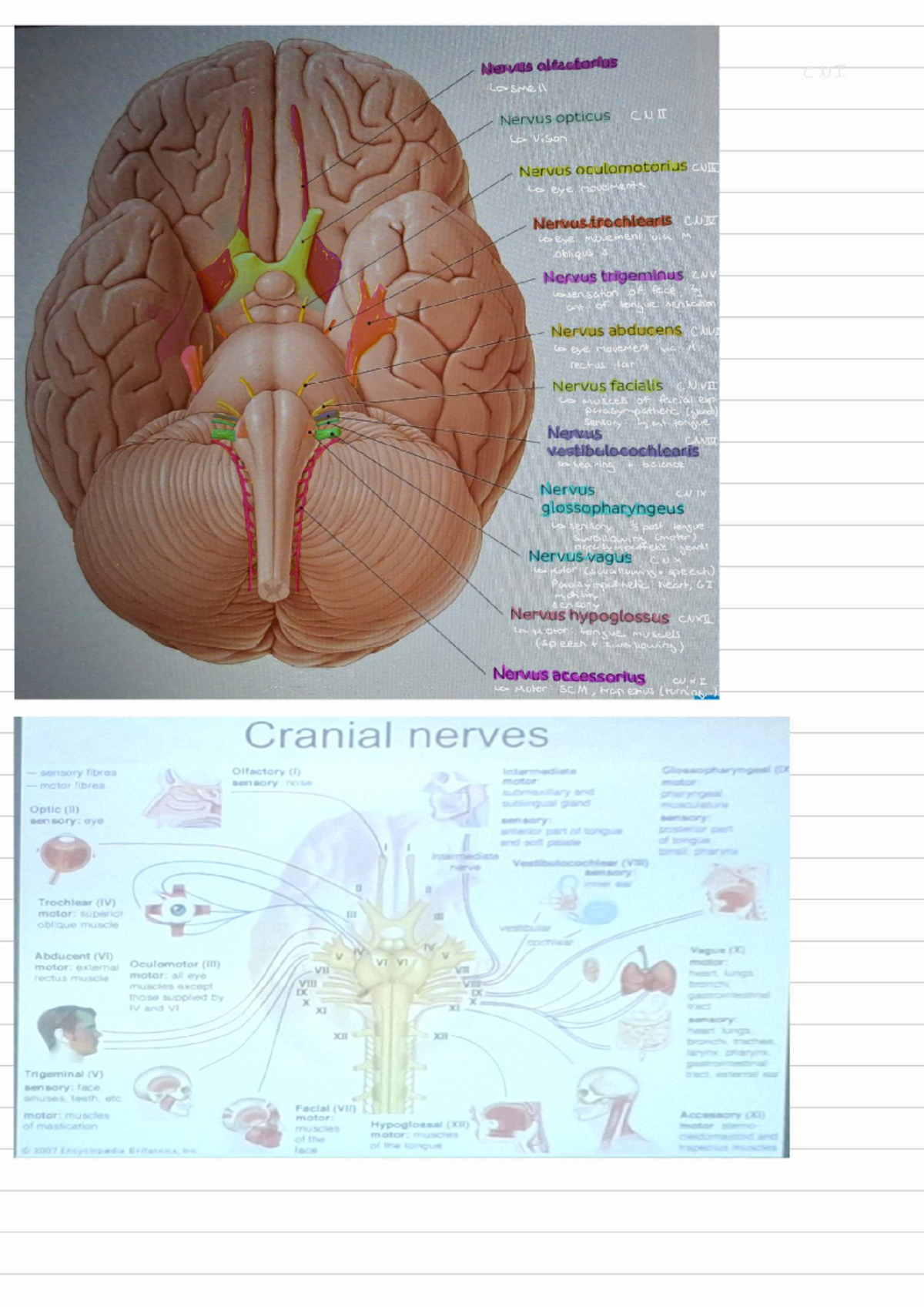
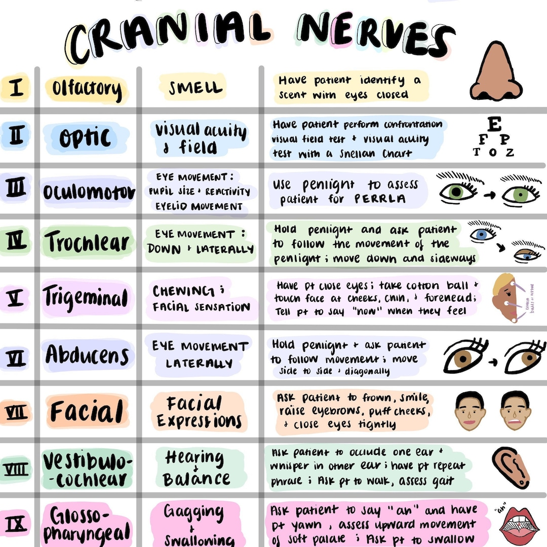
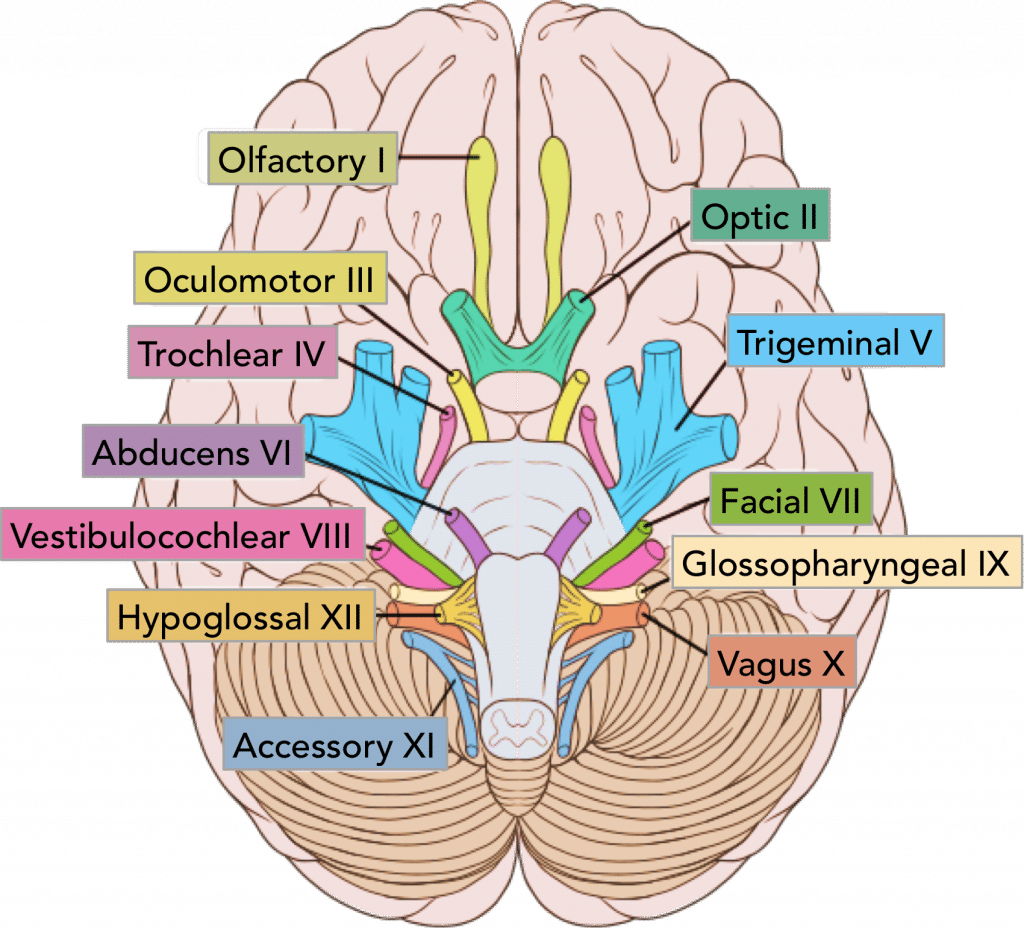
Cranial nerves are a set of 12 paired nerves that arise directly from the brain or brainstem, rather than the spinal cord. They serve essential sensory and motor functions for the head and neck, including sight, smell, hearing, and facial movement, while also regulating autonomic functions like heart rate and digestion.
Understanding the Architecture of Cranial Nerves
Cranial nerves represent a critical component of the peripheral nervous system (PNS), though the first two (olfactory and optic) are technically extensions of the central nervous system. Unlike spinal nerves, which emerge from the spinal cord segments, cranial nerves emerge from the cranium through various openings called foramina. They are traditionally numbered using Roman numerals (I–XII) based on their anatomical position from anterior to posterior (front to back) as they exit the brain.
The functional classification of these nerves is divided into three categories: sensory, motor, or mixed (containing both sensory and motor fibers). Sensory nerves carry impulses from peripheral receptors to the brain, while motor nerves carry impulses from the brain to muscles or glands. Mixed nerves perform both functions. In modern clinical practice in 2026, understanding these pathways remains the foundation for neurological diagnostics, allowing practitioners to pinpoint lesions within the brainstem or specific cranial compartments based on functional deficits.
The Origin and Exit Points
Most cranial nerves originate in the brainstem, which is composed of the midbrain, pons, and medulla oblongata. The specific locations where these nerves attach to the brain are known as their apparent origins. However, their true origins lie within deep clusters of neurons called nuclei. For example, the oculomotor nerve (CN III) originates in the midbrain, while the vagus nerve (CN X) has nuclei located in the medulla.
After leaving the brain, these nerves must traverse the skull to reach their targets. They pass through specific channels in the sphenoid, ethmoid, temporal, and occipital bones. Understanding these exit points, such as the superior orbital fissure or the jugular foramen, is vital for surgeons and neurologists when evaluating traumatic brain injuries or skull base tumors.
Detailed Breakdown of the 12 Cranial Nerves
CN I: The Olfactory Nerve (Sensory)
The olfactory nerve is the first and shortest cranial nerve. Its primary function is the sense of smell (olfaction). Unlike most other nerves, it does not join with the brainstem; instead, it enters the olfactory bulbs, which are extensions of the forebrain.
The nerve fibers pass through the cribriform plate of the ethmoid bone. Damage to this nerve, often caused by head trauma or viral infections, results in anosmia (the loss of smell). Interestingly, because smell is closely linked to taste, patients with CN I damage often complain that food has lost its flavor.
CN II: The Optic Nerve (Sensory)
The optic nerve is responsible for transmitting visual information from the retina to the brain. It is considered an outgrowth of the diencephalon. The two optic nerves meet at the optic chiasm, where some fibers cross to the opposite side of the brain, allowing for binocular vision and depth perception.
Clinical assessment of CN II involves testing visual acuity (Snellen chart), visual fields, and the pupillary light reflex. Conditions such as glaucoma or optic neuritis directly impact this nerve's ability to transmit electrical signals to the primary visual cortex in the occipital lobe.
CN III: The Oculomotor Nerve (Motor)
The oculomotor nerve is the primary "workhorse" for eye movement. It innervates four of the six extraocular muscles: the superior rectus, inferior rectus, medial rectus, and inferior oblique. Additionally, it controls the levator palpebrae superioris, which lifts the upper eyelid.
Beyond skeletal muscle control, CN III provides parasympathetic innervation to the ciliary muscle (for lens accommodation) and the sphincter pupillae (for pupil constriction). A complete oculomotor palsy typically presents as "down and out" eye positioning and a drooping eyelid (ptosis).
CN IV: The Trochlear Nerve (Motor)
The trochlear nerve is unique for two reasons: it is the smallest cranial nerve by fiber count and the only one that emerges from the dorsal (posterior) aspect of the brainstem. It innervates a single muscle: the superior oblique muscle of the eye.
The superior oblique muscle is responsible for rotating the eye downward and inward (intorsion). Damage to CN IV often leads to vertical diplopia (double vision), which is most noticeable when a patient attempts to look down, such as when walking down stairs or reading.
CN V: The Trigeminal Nerve (Mixed)
The trigeminal nerve is the largest cranial nerve and is primarily responsible for facial sensation and the motor functions of chewing (mastication). It divides into three major branches:
- Ophthalmic (V1): Purely sensory, covering the forehead, upper eyelid, and cornea.
- Maxillary (V2): Purely sensory, covering the mid-face, upper lip, and upper teeth.
- Mandibular (V3): Mixed; provides sensation to the lower face and jaw, and motor control to the muscles of mastication (masseter, temporalis).
The trigeminal nerve is the site of trigeminal neuralgia, a chronic pain condition characterized by severe, shock-like facial pain triggered by minor stimuli like touching the face or brushing teeth.
CN VI: The Abducens Nerve (Motor)
The abducens nerve innervates the lateral rectus muscle, which is responsible for abducting the eye (moving it away from the midline). This nerve has a long intracranial course, making it particularly vulnerable to increases in intracranial pressure.
If the abducens nerve is compromised, the patient will experience medial strabismus (the eye turns inward) because the medial rectus muscle (controlled by CN III) is unopposed.
CN VII: The Facial Nerve (Mixed)
The facial nerve is highly complex, controlling the muscles of facial expression, providing taste sensation to the anterior two-thirds of the tongue, and stimulating various glands (lacrimal, submandibular, and sublingual).
A common pathology associated with this nerve is Bell's Palsy, an idiopathic unilateral facial paralysis. Patients lose the ability to close their eye, smile, or frown on the affected side. Because it also controls the stapedius muscle in the ear, damage can lead to hyperacusis (increased sensitivity to sound).
CN VIII: The Vestibulocochlear Nerve (Sensory)
Formerly known as the auditory nerve, CN VIII consists of two distinct components: the vestibular nerve (balance and equilibrium) and the cochlear nerve (hearing). It transmits signals from the inner ear to the brainstem.
Symptoms of vestibulocochlear dysfunction include vertigo, nystagmus (involuntary eye movement), and sensorineural hearing loss. Testing often includes the Weber and Rinne tests or caloric reflex testing.
CN IX: The Glossopharyngeal Nerve (Mixed)
This nerve manages several diverse functions. It provides motor control to the stylopharyngeus muscle (used in swallowing), carries taste and general sensation from the posterior one-third of the tongue, and provides parasympathetic innervation to the parotid salivary gland.
Crucially, CN IX also carries sensory information from the carotid sinus (baroreceptors for blood pressure) and carotid body (chemoreceptors for oxygen/carbon dioxide levels), making it essential for cardiovascular and respiratory homeostasis.
CN X: The Vagus Nerve (Mixed)
The vagus nerve is the longest cranial nerve, extending from the brainstem down into the abdomen. Its name is derived from the Latin word for "wandering." It provides the primary parasympathetic supply to the heart, lungs, and most of the digestive tract.
In addition to its autonomic roles, it controls the muscles of the larynx and pharynx, which are vital for speech and the gag reflex. Vagus nerve stimulation (VNS) has become a significant therapeutic area for treating epilepsy and treatment-resistant depression.
CN XI: The Accessory Nerve (Motor)
The accessory nerve is primarily a motor nerve that controls the sternocleidomastoid and trapezius muscles. These muscles allow for head rotation and shrugging of the shoulders.
Unlike other cranial nerves, the accessory nerve has a "spinal" component that arises from the upper cervical spinal cord, enters the skull through the foramen magnum, and then exits through the jugular foramen. Evaluation involves testing the strength of a patient's shoulder shrug against resistance.
CN XII: The Hypoglossal Nerve (Motor)
The hypoglossal nerve provides motor innervation to all intrinsic and most extrinsic muscles of the tongue. Its function is essential for speech, food manipulation, and swallowing.
If the hypoglossal nerve is damaged on one side, the tongue will deviate toward the side of the injury when the patient sticks it out. This occurs because the healthy genioglossus muscle on the opposite side is stronger and pushes the tongue toward the paralyzed side.
Comparative Summary of Cranial Nerves
The following table summarizes the primary characteristics of the twelve cranial nerves.
| Number | Name | Type | Primary Function | Exit Foramen |
|---|---|---|---|---|
| I | Olfactory | Sensory | Sense of smell | Cribriform plate |
| II | Optic | Sensory | Vision | Optic canal |
| III | Oculomotor | Motor | Eye movement, pupil constriction | Superior orbital fissure |
| IV | Trochlear | Motor | Eye movement (Superior oblique) | Superior orbital fissure |
| V | Trigeminal | Mixed | Facial sensation, mastication | Var. (SOF, Rotundum, Ovale) |
| VI | Abducens | Motor | Eye movement (Lateral rectus) | Superior orbital fissure |
| VII | Facial | Mixed | Facial expression, taste, lacrimation | Internal acoustic meatus |
| VIII | Vestibulocochlear | Sensory | Hearing and balance | Internal acoustic meatus |
| IX | Glossopharyngeal | Mixed | Taste, swallowing, BP monitoring | Jugular foramen |
| X | Vagus | Mixed | Autonomic (heart, GI), swallowing | Jugular foramen |
| XI | Accessory | Motor | Shoulder shrug, head rotation | Jugular foramen |
| XII | Hypoglossal | Motor | Tongue movement | Hypoglossal canal |
Clinical Examination of Cranial Nerves
A thorough neurological exam must systematically evaluate each of these nerves. This is often the first step in diagnosing conditions like multiple sclerosis, brainstem strokes, or localized tumors.
- Olfactory (I): Use non-irritating scents like coffee or soap to test each nostril.
- Optic (II): Check acuity with a chart and peripheral vision via "confrontation."
- Oculomotor, Trochlear, Abducens (III, IV, VI): Follow a finger in an "H" pattern to check eye movement and use a penlight for pupillary response.
- Trigeminal (V): Test facial touch in all three zones and ask the patient to clench their teeth.
- Facial (VII): Ask the patient to smile, frown, and puff out their cheeks.
- Vestibulocochlear (VIII): Use a whisper test or tuning forks to check hearing.
- Glossopharyngeal & Vagus (IX, X): Check for a symmetrical "ah" sound or a gag reflex.
- Accessory (XI): Test shoulder shrug and neck turning resistance.
- Hypoglossal (XII): Inspect the tongue for atrophy or deviation.
Functional Grouping for Easier Recall
To help categorize these nerves logically, clinicians often group them by their primary functional systems rather than just their numerical order.
Special Sensory Nerves
CN I, II, and VIII are purely sensory and deal with the special senses: smell, sight, and hearing/balance. These do not have motor components and are often the focus of specialized assessments in ophthalmology and otolaryngology.
Eye Movement Nerves
CN III, IV, and VI work in tandem to coordinate ocular motility. Their nuclei are connected by the medial longitudinal fasciculus (MLF), a tract in the brainstem that allows both eyes to move together smoothly. Dysfunction in this grouping can lead to nystagmus or "lazy eye" symptoms.
Pharyngeal Arch Nerves
CN V, VII, IX, and X are associated with the embryological pharyngeal arches. They share a mixed nature, providing both sensory innervation to the skin or mucosa and motor control to the muscles derived from these arches, such as the muscles of facial expression (Arch 2) or muscles of swallowing (Arches 4 and 6).
Frequently Asked Questions
Which cranial nerve is the longest?
The vagus nerve (CN X) is the longest of the cranial nerves. Unlike the others that stay mostly within the head and neck, the vagus nerve travels from the medulla down through the neck into the thorax and abdomen, reaching the heart, lungs, and the majority of the digestive system.
How can I remember the order of the cranial nerves?
Many students use mnemonics to remember the order. A classic one is: "On Occasion Our Trusty Truck Acts Funny, Very Good Vehicle Any How." The first letter of each word corresponds to the first letter of the nerves: Olfactory, Optic, Oculomotor, Trochlear, Trigeminal, Abducens, Facial, Vestibulocochlear, Glossopharyngeal, Vagus, Accessory, and Hypoglossal.
What is the "Nerve 0" or the Terminal Nerve?
While the 12 pairs are the standard taught in anatomy, there is a "Nerve 0" known as the terminal nerve. It is a tiny plexus of nerves found anterior to the olfactory nerve. In humans, its function is still debated, though it is thought to be involved in sensing pheromones or regulating reproductive hormones via the gonadotropin-releasing hormone (GnRH) pathway.
Which nerves are involved in the gag reflex?
The gag reflex is primarily a joint effort between the glossopharyngeal nerve (CN IX) and the vagus nerve (CN X). CN IX provides the sensory limb (feeling the stimulus in the back of the throat), while CN X provides the motor limb (the muscular contraction that causes the "gag").
Can cranial nerves regenerate after damage?
Regeneration depends on the severity and type of injury. Because they are part of the peripheral nervous system, some cranial nerves have the potential for slow regeneration if the cell body is intact and the myelin sheath provides a pathway. However, nerves that are extensions of the CNS, like the optic nerve, generally do not regenerate effectively once damaged.
Practical Next Actions
If you are studying for medical or biological examinations, the next logical step is to practice a hands-on neurological exam. Understanding the theory of "what each nerve does" is significantly reinforced by observing the "how" of clinical testing. Reviewing high-quality anatomical atlases, such as those provided by the National Library of Medicine or peer-reviewed platforms like StatPearls, can provide deeper insights into the specific nuclei and pathways mentioned in this guide. For those experiencing symptoms related to facial paralysis, vision loss, or hearing changes, consulting a board-certified neurologist is the recommended course of action for a definitive diagnosis.