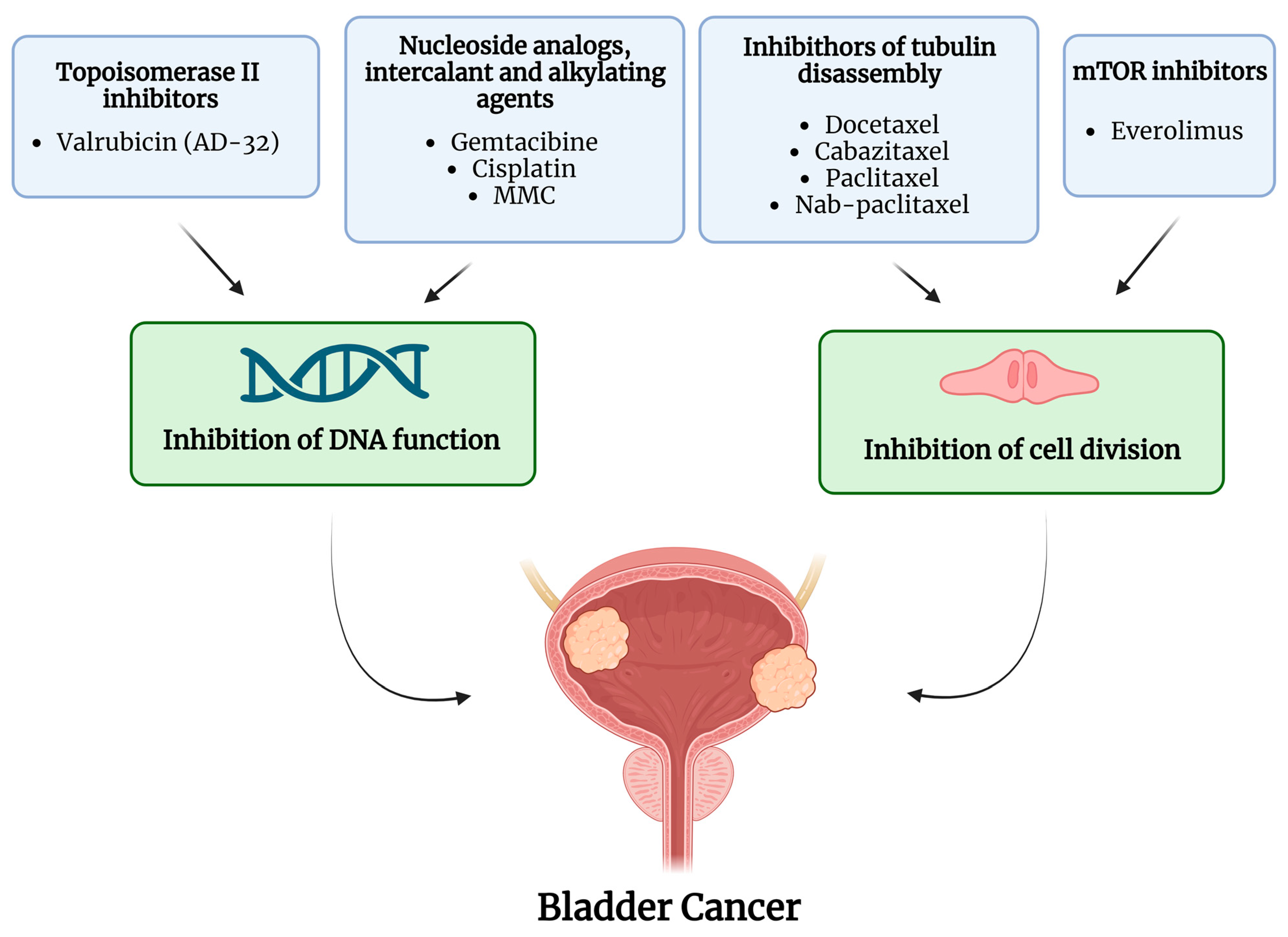
T4 bladder cancer represents the most advanced local stage of urothelial carcinoma, defined by the tumor invading tissues immediately surrounding the bladder, such as the prostate, uterus, vagina, pelvic wall, or abdominal wall. Managing this diagnosis requires a comprehensive understanding of staging metrics, multidisciplinary treatment combinations including chemotherapy and radical surgery, and supportive care options aimed at optimizing patient outcomes and quality of life.
Defining T4 Bladder Cancer and the TNM Staging System
Understanding a T4 bladder cancer diagnosis requires looking closely at how oncologists classify cancer spread using the international TNM staging system. Maintained by organizations such as the American Joint Committee on Cancer (AJCC) and the Union for International Cancer Control (UICC), this framework evaluates the primary tumor (T), regional lymph nodes (N), and distant metastasis (M).
In this framework, the "T" category describes how deeply the tumor has grown through the muscular wall of the bladder into surrounding tissues.
- Ta and Tis: Non-invasive papillary carcinoma and carcinoma in situ (CIS), confined to the inner epithelial lining.
- T1: The tumor has grown through the lining into the connective tissue beneath (lamina propria).
- T2: The tumor has invaded the muscle layer (detrusor muscle).
- T3: The tumor has grown past the muscle into the surrounding perivesical fatty tissue.
- T4: The tumor has grown outside the bladder into adjacent pelvic organs or the pelvic/abdominal wall.
Sub-Classification of T4 Bladder Cancer
The T4 category is further split into two distinct sub-stages depending on the exact anatomical structures invaded by the primary tumor:
- T4a: The tumor has invaded the prostate stroma, seminal vesicles, uterus, or vagina. In individuals assigned male at birth, this indicates invasion into the muscular and connective tissue framework of the prostate. In individuals assigned female at birth, it involves the uterine corpus, cervix, or vaginal wall.
- T4b: The tumor has invaded the pelvic wall or the abdominal wall. This stage often indicates that the cancer is fixed to the bony pelvis or musculature of the lower abdomen, which significantly alters surgical resectability and treatment planning.
Distinguishing between T4a and T4b is crucial because it directly influences whether a patient is a candidate for radical extirpative surgery or whether treatment must focus primarily on bladder-preservation protocols, systemic therapies, and palliative interventions.
Diagnostic Workup and Staging Evaluations
Accurately staging a patient suspected of having T4 bladder cancer demands a rigorous, multi-modal diagnostic workup. Because treatment strategies carry significant life-altering implications, clinical teams must precisely map the boundaries of the tumor and rule out systemic spread.
Clinical Evaluation and Imaging
The diagnostic journey typically begins with a detailed physical examination, including a bimanual examination performed under anesthesia to assess the mobility or fixation of the bladder and whether a palpable mass extends into the pelvic sidewall.
Advanced cross-sectional imaging is mandatory to visualize local invasion and distant metastases:
- Computed Tomography (CT) Urogram: Provides detailed cross-sectional views of the kidneys, ureters, and bladder, helping identify local soft-tissue extension and retroperitoneal lymph node enlargement.
- Magnetic Resonance Imaging (MRI) of the Pelvis: Offers superior soft-tissue contrast compared to CT, making it exceptionally useful for distinguishing T4a prostatic or vaginal invasion from lesser stages of muscle-invasive disease.
- Chest Imaging: Chest CT scans are routinely utilized to screen for pulmonary metastases, which represent a common site of distant spread for urothelial carcinoma.
- Bone Scans or Positron Emission Tomography (PET-CT): Employed when clinical symptoms or laboratory findings suggest potential skeletal involvement.
Transurethral Resection of the Bladder Tumor (TURBT)
While imaging provides a macroscopic view, histopathological confirmation via TURBT remains the gold standard. During a TURBT, a urologic surgeon uses a cystoscope to resect visible tumor tissue and deep muscle biopsies from the base of the lesion. Pathologists examine these specimens to confirm the histological subtype—most commonly urothelial carcinoma, though variants such as micropapillary, nested, or plasmacytoid variants can influence aggressiveness—and to confirm the depth of detrusor muscle and perivesical tissue penetration.
Comparing Treatment Modalities for T4 Disease
Management of T4 bladder cancer requires a coordinated multidisciplinary team comprising urologic oncologists, medical oncologists, radiation oncologists, pathologists, and specialized oncology nurses. The table below outlines the primary treatment modalities, their mechanisms, and their standard clinical goals.
| Treatment Modality | Primary Mechanism | Clinical Goal | Key Considerations |
|---|---|---|---|
| Neoadjuvant Chemotherapy | Systemic administration of cytotoxic drugs prior to local therapy. | Eradicate micrometastases and downstage the primary tumor. | Platinum-based regimens (e.g., cisplatin/gemcitabine) are standard for eligible patients. |
| Radical Cystectomy & Urinary Diversion | Surgical removal of the bladder, nearby organs, and regional lymph nodes. | Achieve local control and potential long-term survival. | Often combined with urinary diversion (e.g., ileal conduit or neobladder). |
| Trimodality Therapy (Bladder Preservation) | Combination of maximal TURBT, concurrent chemotherapy, and external beam radiation. | Preserve native bladder function while controlling local disease. | Preferred for patients unfit for surgery or those prioritizing organ preservation. |
| Systemic Immunotherapy | Immune checkpoint inhibition (e.g., PD-1/PD-L1 inhibitors). | Harness the patient's immune system to target cancer cells. | Used in cisplatin-ineligible settings, post-platinum progression, or maintenance therapy. |
Systemic Therapies and Chemotherapy Protocols
Because T4 bladder cancer is an advanced local disease, microscopic dissemination outside the primary site is common. Consequently, systemic therapy forms the backbone of modern management strategies.
Platinum-Based Combination Chemotherapy
For patients with adequate renal function and performance status, cisplatin-based combination chemotherapy is the standard first-line approach. The two most widely validated regimens include:
- GC (Gemcitabine and Cisplatin): Generally favored due to a more favorable toxicity profile compared to older regimens, while maintaining high efficacy.
- dd-MVAC (Dose-Dense Methotrexate, Vinblastine, Doxorubicin, and Cisplatin): Delivered with granulocyte colony-stimulating factor (G-CSF) support to shorten cycle intervals, demonstrating high pathological complete response rates.
For patients who are ineligible for cisplatin—often due to impaired renal function, peripheral neuropathy, or poor performance status—carboplatin-based regimens or alternative non-platinum options are evaluated on an individual basis.
The Role of Immunotherapy and Targeted Agents
The therapeutic landscape for advanced urothelial carcinoma has expanded significantly with the integration of immune checkpoint inhibitors. Drugs targeting the programmed cell death protein 1 (PD-1) or its ligand (PD-L1) offer durable responses in a subset of patients. Furthermore, antibody-drug conjugates (ADCs) and targeted therapies directed at specific genetic alterations, such as fibroblast growth factor receptor (FGFR) mutations, provide additional lines of defense when standard chemotherapy proves insufficient.
Local Therapy: Surgery Versus Bladder Preservation
Deciding between radical surgery and bladder-preservation protocols in T4 disease is complex and depends heavily on whether the tumor is classified as T4a or T4b, alongside patient fitness and personal preferences.
Radical Cystectomy and Pelvic Lymph Node Dissection
For carefully selected patients with T4a disease—particularly where local invasion is limited and can be excised with negative margins—radical cystectomy offers the highest likelihood of long-term local disease control.
- In individuals assigned male at birth, this procedure typically involves the removal of the bladder, prostate, and seminal vesicles.
- In individuals assigned female at birth, it involves the removal of the bladder, uterus, ovaries, and anterior wall of the vagina.
A comprehensive pelvic lymph node dissection is performed concurrently to stage and remove potentially involved nodal basins. Following cystectomy, surgeons construct a form of urinary diversion, ranging from an incontinent ileal conduit to a continent cutaneous reservoir or an orthotopic neobladder, depending on anatomical and oncological constraints.
Multimodality Bladder-Preservation Therapy
When a tumor is fixed to the pelvic wall (T4b) or when a patient has significant comorbidities that preclude major pelvic surgery, trimodality therapy serves as a viable alternative. This approach begins with a maximal transurethral resection of the bladder tumor to reduce tumor burden, followed by concurrent chemoradiation therapy. Radiation is delivered to the bladder and regional lymph nodes simultaneously with radiosensitizing chemotherapy agents, allowing many patients to retain native bladder function without compromising survival outcomes.
Prognosis, Surveillance, and Survivorship
The prognosis for T4 bladder cancer is guarded, reflecting the advanced nature of local invasion and the risk of metastatic spread. Survival statistics vary widely based on patient age, baseline health, exact anatomical substage (T4a versus T4b), regional lymph node involvement, and response to systemic therapy.
Following the completion of primary treatment, rigorous surveillance protocols are essential. Routine follow-up typically involves:
- Regular cross-sectional imaging (CT or MRI) of the chest, abdomen, and pelvis.
- Periodic cystoscopic evaluations if the bladder was preserved.
- Monitoring of renal function, electrolyte balances, and metabolic status, particularly for patients with urinary diversions.
- Assessment and management of long-term toxicities related to pelvic radiation, chemotherapy, or surgical alterations.
Supportive care—including nutritional counseling, management of urinary diversion stomas or urinary symptoms, physical rehabilitation, and psycho-oncology support—plays an integral role throughout the survivorship continuum.
Disclaimer: This overview is intended for informational and educational purposes only and should not be construed as medical advice. Any diagnosis of T4 bladder cancer requires personalized evaluation, treatment planning, and management by qualified oncological specialists.