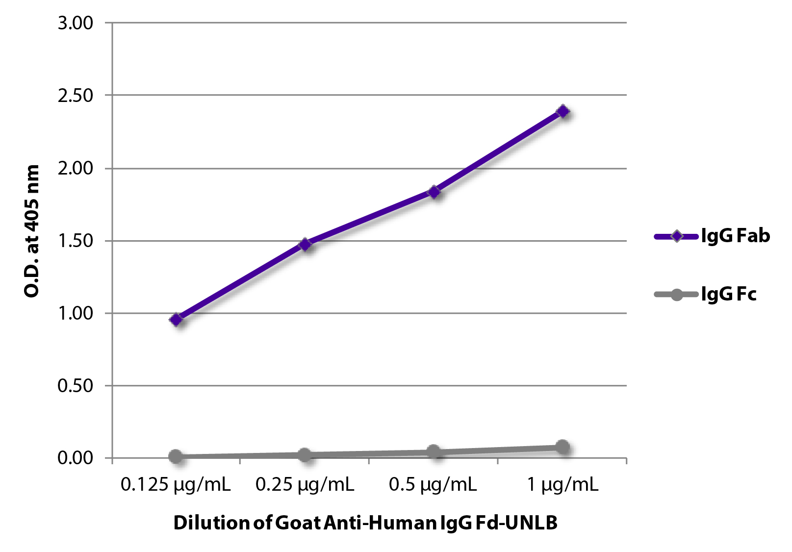
Anti-human IgG Fc antibodies are specialized secondary antibodies designed to bind specifically to the Fragment crystallizable (Fc) region of human Immunoglobulin G (IgG). In 2026, they remain fundamental tools in research and clinical diagnostics, used to detect, quantify, or capture human IgG without interfering with the antibody's antigen-binding sites (Fab).
The ability to target the Fc region specifically is essential for assays where maintaining the structural integrity of the primary antibody-antigen complex is required. By binding to the heavy chain constant regions (CH2 and CH3 domains), these secondary antibodies provide a reliable signal in applications ranging from basic enzyme-linked immunosorbent assays (ELISA) to complex therapeutic drug monitoring and pharmacokinetic studies.
Understanding the Molecular Structure: Why Target the Fc Region?
To appreciate the utility of an anti-human IgG Fc antibody, one must first understand the anatomy of the human IgG molecule. IgG is a Y-shaped protein consisting of two heavy chains and two light chains. It is divided into two functional regions:
- The Fab Region (Fragment, antigen-binding): The "arms" of the Y, which contain the variable domains responsible for recognizing and binding to specific antigens.
- The Fc Region (Fragment, crystallizable): The "stem" of the Y, composed of the constant domains of the heavy chains. This region interacts with cell surface receptors (Fc receptors) and the complement system to mediate effector functions.
When researchers use a secondary antibody that targets the Fc region, they ensure that the secondary antibody does not compete with the primary antibody for the antigen-binding site. Furthermore, because the Fc region is highly conserved within a species, an anti-human IgG Fc antibody will recognize almost all human IgG molecules regardless of their specificity for an antigen. This makes it a universal detection tool for human antibodies in various experimental setups.
Specificity vs. Cross-Reactivity
A critical distinction in immunology is the difference between an antibody that targets the "Whole Molecule" (H+L) and one that is "Fc-specific." An antibody targeting the whole molecule reacts with both the heavy (H) and light (L) chains. Since light chains (kappa and lambda) are shared across different classes of immunoglobulins (IgA, IgM, IgE), a "Whole Molecule" secondary antibody may cross-react with these other classes.
In contrast, an anti-human IgG Fc antibody is specifically purified to react only with the heavy chain of IgG. This specificity is vital when the researcher needs to detect only the IgG class in a sample that may also contain human IgM or IgA, such as in serum or plasma samples.
Production and Host Species Selection
As of 2026, the production of anti-human IgG Fc antibodies has reached high levels of refinement. These antibodies are typically produced by immunizing a non-human host species with purified human IgG Fc fragments.
Common Host Species
- Goat: The most common host for polyclonal antibodies due to the large volume of serum that can be collected and the strong immune response goats mount against human proteins.
- Rabbit: Often used for generating high-affinity monoclonal or polyclonal antibodies. Rabbit secondaries are frequently preferred for Western blotting due to their clean signaling.
- Donkey: Often chosen for multiple labeling experiments. If a researcher is using multiple primary antibodies from different species (e.g., mouse, rat, and human), using donkey-derived secondaries that have been cross-adsorbed against those other species minimizes background noise.
- Mouse: Primarily used for generating monoclonal anti-human IgG Fc antibodies, which offer high batch-to-batch consistency and exquisite specificity for particular IgG subclasses (IgG1, IgG2, IgG3, or IgG4).
Polyclonal vs. Monoclonal Secondaries
Polyclonal anti-human IgG Fc antibodies are a mixture of antibodies that recognize multiple epitopes on the Fc fragment. This "multi-target" binding leads to signal amplification, making them highly sensitive for detecting low-abundance targets.
Monoclonal antibodies, however, recognize a single epitope. While they might provide a slightly lower signal intensity, they offer superior specificity and are essential in standardized clinical diagnostic kits where reproducibility over decades is required.
The Importance of Cross-Adsorption (Pre-adsorption)
In many laboratory environments, samples are complex. For example, if a researcher is performing an immunohistochemistry (IHC) stain on mouse tissue using a human primary antibody, they must use an anti-human IgG Fc secondary. However, because human and mouse IgG share structural similarities, the anti-human secondary might "accidentally" bind to the endogenous mouse IgG in the tissue, leading to high background staining.
To prevent this, manufacturers use cross-adsorption. This process involves passing the anti-human IgG Fc antibody through a column containing immobilized proteins from other species (such as mouse, rat, or bovine serum). Antibodies that cross-react bind to the column and are removed, while the highly specific anti-human antibodies pass through. In 2026, "highly cross-adsorbed" antibodies are the gold standard for multiplexed assays and tissue staining.
Conjugation and Detection Methods
The anti-human IgG Fc antibody itself is invisible; it must be conjugated to a "reporter" molecule to be detected. The choice of conjugate depends on the specific application and the required sensitivity.
Enzymatic Conjugates (HRP and AP)
- Horseradish Peroxidase (HRP): The most popular enzyme for Western blotting and ELISA. It is small, stable, and reacts with substrates (like TMB or ECL) to produce a color change or light (chemiluminescence).
- Alkaline Phosphatase (AP): Often used in ELISAs and IHC. It is generally considered to have higher sensitivity than HRP in certain colorimetric assays but has a slower reaction rate.
Fluorescent Conjugates (Fluorophores)
With the advancement of imaging in 2026, fluorescently labeled anti-human IgG Fc antibodies are ubiquitous.
- Classic Dyes: FITC (Green) and TRITC (Red).
- Modern Dyes: Alexa Fluor® dyes, Cyanine (Cy3/Cy5), and DyLight® fluorophores. These offer superior brightness, photostability, and pH resistance, making them ideal for confocal microscopy and flow cytometry.
Biotinylation
Biotinylated anti-human IgG Fc antibodies are used in conjunction with Streptavidin-labeled probes. Because one Streptavidin molecule can bind four Biotin molecules, this creates an amplification cascade, significantly increasing the signal for very low-concentration targets.
Primary Applications in Research and Diagnostics
1. ELISA (Enzyme-Linked Immunosorbent Assay)
In an indirect ELISA, a specific antigen is coated on a plate. The human primary antibody (e.g., from a patient's serum) binds to the antigen. The anti-human IgG Fc-HRP is then added to detect the bound human IgG. This is the standard method for testing for viral exposure (like HIV or SARS-CoV variants) or autoimmune disorders.
2. Western Blotting
Researchers use anti-human IgG Fc to detect human antibodies or Fc-fusion proteins that have been separated by gel electrophoresis. Using an Fc-specific secondary ensures that the heavy chain (approx. 50 kDa) is detected without interference from the light chain (approx. 25 kDa), which is useful if the target protein has a molecular weight similar to the light chain.
3. Therapeutic Antibody Development (Pharmacokinetics)
Most modern monoclonal antibody drugs (mAbs) are human or humanized IgG. To study how long these drugs stay in a patient's or animal's system, researchers use anti-human IgG Fc antibodies to capture and quantify the drug in serum samples without detecting the host's own non-human antibodies.
4. Flow Cytometry
In flow cytometry, fluorescently labeled anti-human IgG Fc is used to detect human antibodies bound to cell surface markers. This is critical in immunology for characterizing cell populations in humanized mouse models.
Comparison of Secondary Antibody Formats
The following table compares the different formats of anti-human IgG Fc antibodies available for laboratory use in 2026.
| Antibody Format | Best Use Case | Advantages | Limitations |
|---|---|---|---|
| Polyclonal HRP-Conjugated | Standard ELISA, Western Blot | High sensitivity, cost-effective | Batch-to-batch variability |
| Monoclonal (Clone-specific) | Clinical diagnostics, Subclassing | Exceptional reproducibility | Lower signal amplification |
| Highly Cross-Adsorbed | Multiplex IHC, Tissue staining | Low background in complex samples | Higher cost per mg |
| F(ab')2 Fragment | Cell-based assays (Flow Cytometry) | Prevents binding to host Fc receptors | More difficult to manufacture |
| Biotinylated | Low-abundance target detection | Significant signal amplification | Requires extra incubation step |
Technical Selection Criteria: Choosing the Right Antibody
When selecting an anti-human IgG Fc antibody in 2026, researchers should follow a structured decision-making process to ensure experimental success:
- Determine the Class/Subclass: Do you need to detect all human IgG, or a specific subclass like IgG1? If the latter, a monoclonal antibody is required.
- Assess the Sample Source: If your sample contains other species' proteins (e.g., a mouse tissue section), you must choose a secondary that has been cross-adsorbed against that specific species.
- Choose the Reporter: Use HRP for chemiluminescence (Western Blot) or fluorophores with non-overlapping spectra for multi-color microscopy.
- Consider Fragment Antibodies: In some cases, using a whole IgG secondary antibody can cause "background" because the secondary antibody's own Fc region binds to Fc receptors on the cells being studied. In these instances, using a F(ab')2 fragment of the anti-human IgG Fc antibody (where the secondary's Fc region has been enzymatically removed) is recommended.
Troubleshooting Common Issues
High Background Noise
If the entire assay shows a signal regardless of the presence of the target, the anti-human IgG Fc may be too concentrated, or it may be cross-reacting with other proteins.
- Solution: Increase the dilution of the secondary antibody or use a cross-adsorbed version. Ensure blocking buffers (like BSA or Normal Serum) are used appropriately.
Weak or No Signal
This often occurs if the secondary antibody does not recognize the primary antibody, or if the enzyme conjugate has lost activity.
- Solution: Verify the primary antibody is indeed a human IgG. Check the expiration of the HRP or fluorescent conjugate. Ensure that no sodium azide was used in HRP-based buffers, as azide irreversibly inhibits HRP.
Cross-Reactivity with Other Immunoglobulins
If you are trying to detect IgG but are getting signals for IgM as well, your antibody is likely not Fc-specific.
- Solution: Switch to an antibody explicitly labeled as "Fc-specific" rather than "(H+L)".
Frequently Asked Questions
What is the difference between Anti-Human IgG Fc and Anti-Human IgG (H+L)?
Anti-human IgG (H+L) reacts with both the heavy and light chains of the IgG molecule. Because light chains are shared with IgM and IgA, (H+L) antibodies are less specific to the IgG class. Anti-human IgG Fc reacts only with the heavy chain constant region, making it specific to the IgG class only.
Can I use an anti-human IgG Fc antibody to detect humanized antibodies in mice?
Yes, this is a common application. However, you must use an anti-human IgG Fc antibody that has been highly cross-adsorbed against mouse IgG to ensure the secondary does not bind to the mouse's own antibodies, which would create massive background noise.
Why is goat often the host species for these antibodies?
Goats are large enough to provide significant quantities of antisera, and their evolutionary distance from humans allows them to produce high-affinity antibodies against human protein structures. Goat anti-human secondaries are the industry standard for general laboratory work.
Does "Fc-specific" mean it will detect all IgG subclasses?
Generally, yes. Most polyclonal anti-human IgG Fc antibodies are designed to recognize common epitopes across IgG1, IgG2, IgG3, and IgG4. If you need to distinguish between these subclasses, you should use a monoclonal antibody specific to the subclass of interest.
Is sodium azide compatible with anti-human IgG Fc-HRP?
No. Sodium azide is a common preservative used to prevent bacterial growth, but it is a potent inhibitor of Horseradish Peroxidase (HRP). If you are using an HRP-conjugated antibody, ensure your wash buffers and antibody diluents are azide-free.
Final Recommendation
For most standard research applications in 2026, a Goat Anti-Human IgG Fc (Polyclonal), HRP-conjugated and Cross-Adsorbed antibody provides the best balance of sensitivity, specificity, and cost. If you are working with multi-color fluorescence or complex tissue samples, prioritize "Highly Cross-Adsorbed" versions to ensure clean, publication-quality results. Always validate the optimal dilution through a titration experiment (checkerboard ELISA) before committing to a large-scale study.