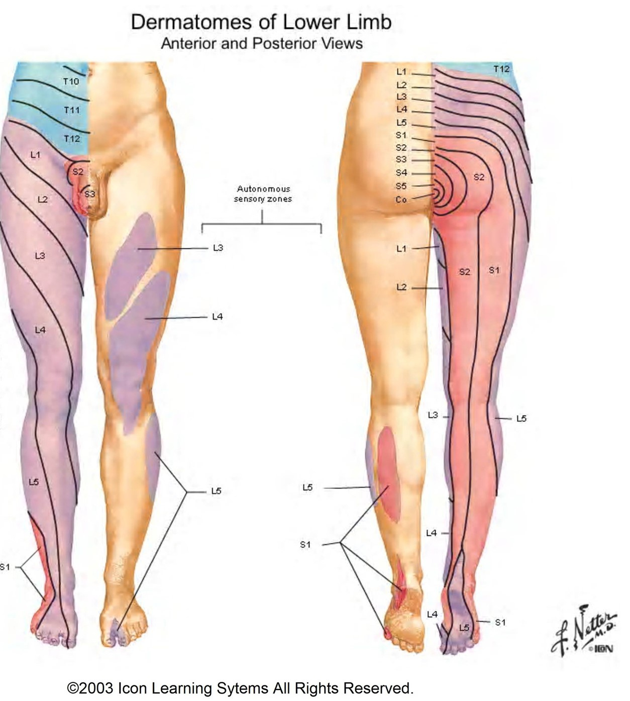
The S1, S2, and S4 dermatomes represent specific areas of skin supplied by sensory neurons originating from the sacral nerve roots of the spinal cord. Clinicians utilize these mapping zones to identify spinal cord injuries, nerve root compression, and peripheral neuropathies, as each segment corresponds to distinct sensory landmarks on the lower limbs and the perineal region.
The Anatomical Significance of Sacral Dermatomes
A dermatome is defined as an area of skin where sensory nerves derive from a single spinal nerve root. In the human body, the sacral region is governed by five pairs of sacral nerves (S1 through S5) exiting the sacrum. These nerves are critical not only for sensation but also for the autonomic functions of the pelvic organs. Understanding the distribution of S1, S2, and S4 is fundamental in neurology and physical medicine because these segments allow practitioners to localize damage within the lower vertebral column or the spinal canal.
When a patient presents with symptoms such as numbness, tingling (paresthesia), or localized pain, mapping these feelings to a specific dermatome allows medical professionals to differentiate between a peripheral nerve injury—which follows the path of a nerve—and a radiculopathy, which follows the distribution of a nerve root exiting the spine.
Mapping the S1 Dermatome: The Lateral Foot and Heel
The S1 dermatome is perhaps the most clinically recognizable of the sacral levels due to its prominent role in physical testing. It covers the lateral aspect of the foot, the lateral malleolus (the outer ankle bone), and extends to the heel.
In clinical practice, the S1 nerve root is frequently evaluated through the ankle-jerk reflex (the Achilles reflex). Diminution or absence of this reflex often indicates pathology at the S1 level. Furthermore, patients with S1 radiculopathy, often caused by a herniated disc at the L5-S1 intervertebral level, typically describe radiating pain traveling down the posterior thigh, through the calf, and settling in the outer edge of the foot or the small toe.
Mapping the S2 Dermatome: The Posterior Thigh and Calf
The S2 dermatome provides sensory coverage for a significant portion of the posterior lower extremity. It occupies the middle region of the posterior thigh and the middle of the calf. While it often overlaps with S1, the S2 dermatome is distinct in its primary focus on the posterior medial aspect of the leg.
When evaluating S2, clinicians look for sensory deficits that do not extend as far down into the foot as S1, but rather remain in the posterior muscular compartments. Damage here is often associated with conditions affecting the cauda equina—the bundle of nerve roots at the end of the spinal cord. Because S2 plays a role in the innervation of the pelvic floor and sexual function, sensory changes in this area are considered high-priority clinical findings that may necessitate urgent diagnostic imaging, such as an MRI.
Mapping the S4 Dermatome: The Perineal and Anogenital Region
The S4 dermatome is vital for its involvement in the "saddle area." Unlike S1 and S2, which primarily govern limb sensation, S4 is concentrated in the perineum, the skin surrounding the anus, and the external genitalia. This dermatome is categorized as part of the sacral parasympathetic outflow, making its integrity essential for bowel, bladder, and sexual function.
The clinical significance of S4 cannot be overstated. A change in sensation in the S4 dermatome—often described as "saddle anesthesia"—is a classic "red flag" symptom in cases of Cauda Equina Syndrome (CES). If a patient experiences loss of sensation in the S4 area alongside bowel or bladder dysfunction, it is widely regarded as a medical emergency requiring rapid intervention to prevent permanent neurological damage.
Comparative Overview of Sacral Dermatomes
| Dermatome | Primary Anatomical Landmark | Clinical Reflex/Association |
|---|---|---|
| S1 | Lateral foot, heel, small toe | Achilles tendon reflex |
| S2 | Posterior thigh, mid-calf | Posterior leg sensation |
| S4 | Perineum, perianal skin, genitalia | Bowel/bladder autonomic control |
Clinical Evaluation and Diagnostic Procedures
To evaluate these dermatomes, a neurologist or physician performs a sensory examination. This typically involves testing light touch and pin-prick sensation across the identified zones. The examination is conducted bilaterally to compare the affected side against the unaffected side.
- Light Touch Testing: Using a cotton wisp or a gloved finger to stroke the skin, the clinician assesses if the patient perceives the sensation as symmetrical.
- Pin-Prick Testing: Using a sterile, disposable safety pin or neurological pinwheel, the clinician tests the patient’s ability to detect sharp stimuli. Diminished sensation in the S4 region during this test is particularly concerning for CES.
- Reflex Testing: Assessing the S1-mediated ankle reflex is standard in orthopedic and neurological screenings.
- Functional Mapping: Because dermatomes have natural overlap, clinicians often use "key points" to distinguish between segments. For example, the S1 area is mapped at the lateral heel, while S2 is mapped at the popliteal fossa (the back of the knee).
Common Pathologies Affecting Sacral Nerves
Several conditions can impact the transmission of sensory information through these dermatomes. Disc herniations are the most common cause of radiculopathy. Specifically, the L5-S1 disc is a frequent site of mechanical compromise, often leading to S1 nerve root irritation.
Spinal stenosis—a narrowing of the spinal canal—can also compress multiple sacral nerve roots simultaneously, leading to bilateral symptoms. In chronic cases, the compression of these nerves may cause "claudication," where pain increases with walking or standing and decreases with sitting, as the spinal canal diameter shifts slightly in different postures.
Differential Diagnosis and Symptom Management
It is important to distinguish between dermatomal pain and other types of sensory disturbance. For instance, peripheral neuropathy, often caused by diabetes, usually follows a "stocking-glove" distribution rather than a specific spinal root pattern. In contrast, nerve root-based symptoms (radiculopathy) typically follow a specific, linear pattern down the leg or toward the perineum.
Patients experiencing these symptoms in 2026 generally undergo a standard diagnostic pathway:
- Physical Examination: Assessing reflex integrity and sensory mapping.
- Electromyography (EMG) and Nerve Conduction Studies: These tests measure electrical activity in the muscles and the speed of nerve signals, helping to verify if a nerve is blocked or damaged.
- Imaging: MRI remains the gold standard for visualizing the relationship between intervertebral discs and sacral nerve roots.
When to Seek Immediate Medical Attention
Because S4 involvement is linked to autonomic function, any sudden change in this specific dermatome requires immediate evaluation. Symptoms that warrant an emergency room visit include:
- Sudden loss of sensation in the inner thighs or perineum ("saddle anesthesia").
- New onset of urinary retention or incontinence.
- Sudden change in bowel function (fecal incontinence).
- Progressive weakness in both legs rather than just one.
These symptoms suggest that the cauda equina is under significant pressure. In such cases, surgery is often necessary to decompress the nerves and preserve long-term pelvic function.
Frequently Asked Questions
What is the difference between S1 and S2 dermatomes? S1 is primarily associated with the lateral foot and the Achilles reflex, while S2 covers the posterior thigh and calf. While they share some territory in the posterior leg, they are distinguished by their specific reflex arcs and their connection to different segments of the sciatic nerve pathway.
Why is S4 considered a red flag in clinical exams? S4 is part of the sacral segment responsible for autonomic control of the bladder and bowel. Sensory loss in the S4 dermatome (the perineal area) often indicates compression of the cauda equina, which can lead to permanent loss of bowel or bladder control if not treated immediately.
Can dermatomes overlap? Yes, dermatomes are not distinct, isolated islands. There is significant overlapping of sensory coverage between adjacent levels. This means that a minor injury to one nerve root might not cause complete numbness because the neighboring nerves provide "collateral" sensory input to that area.
How is a dermatome test performed by a doctor? A doctor will use a series of physical tests, such as touching the skin with a cotton ball to assess light touch or a pin to assess sharp/dull discrimination. They will compare the patient's reaction on the left side to the right side to identify any deficits.
Is it normal to have different sensations in the S1 and S2 areas? It is normal to have uniform sensation across these areas. If one area feels significantly different (numbness, burning, or lack of sensation), it usually indicates nerve root irritation or compression that requires investigation by a specialist.
Conclusion and Next Actions
If you are experiencing symptoms such as unexplained pain, numbness, or tingling in the regions corresponding to the S1, S2, or S4 dermatomes, it is essential to consult with a primary care physician or a neurologist. Document the exact location of the symptoms, whether they fluctuate with movement, and if there are any associated changes in bladder or bowel habits.
If you suspect you have S4 involvement—manifesting as saddle anesthesia or pelvic floor dysfunction—seek urgent medical evaluation. Early identification of nerve root issues significantly improves the likelihood of successful recovery through physical therapy, targeted medication, or appropriate surgical intervention.