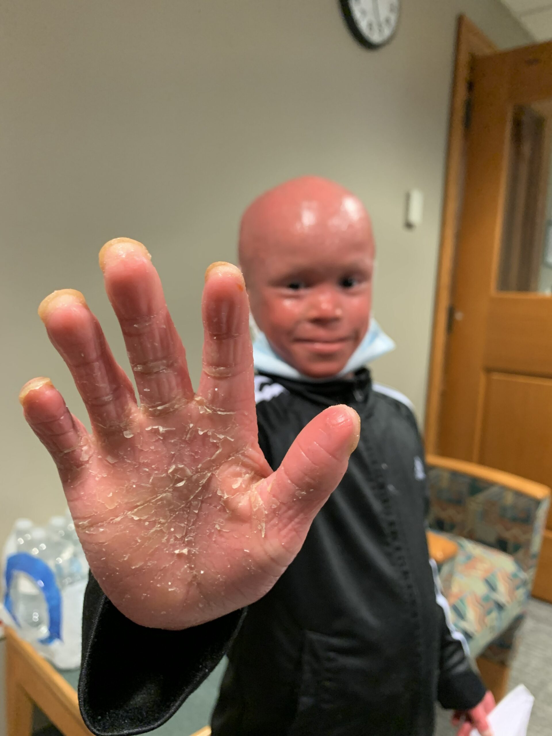
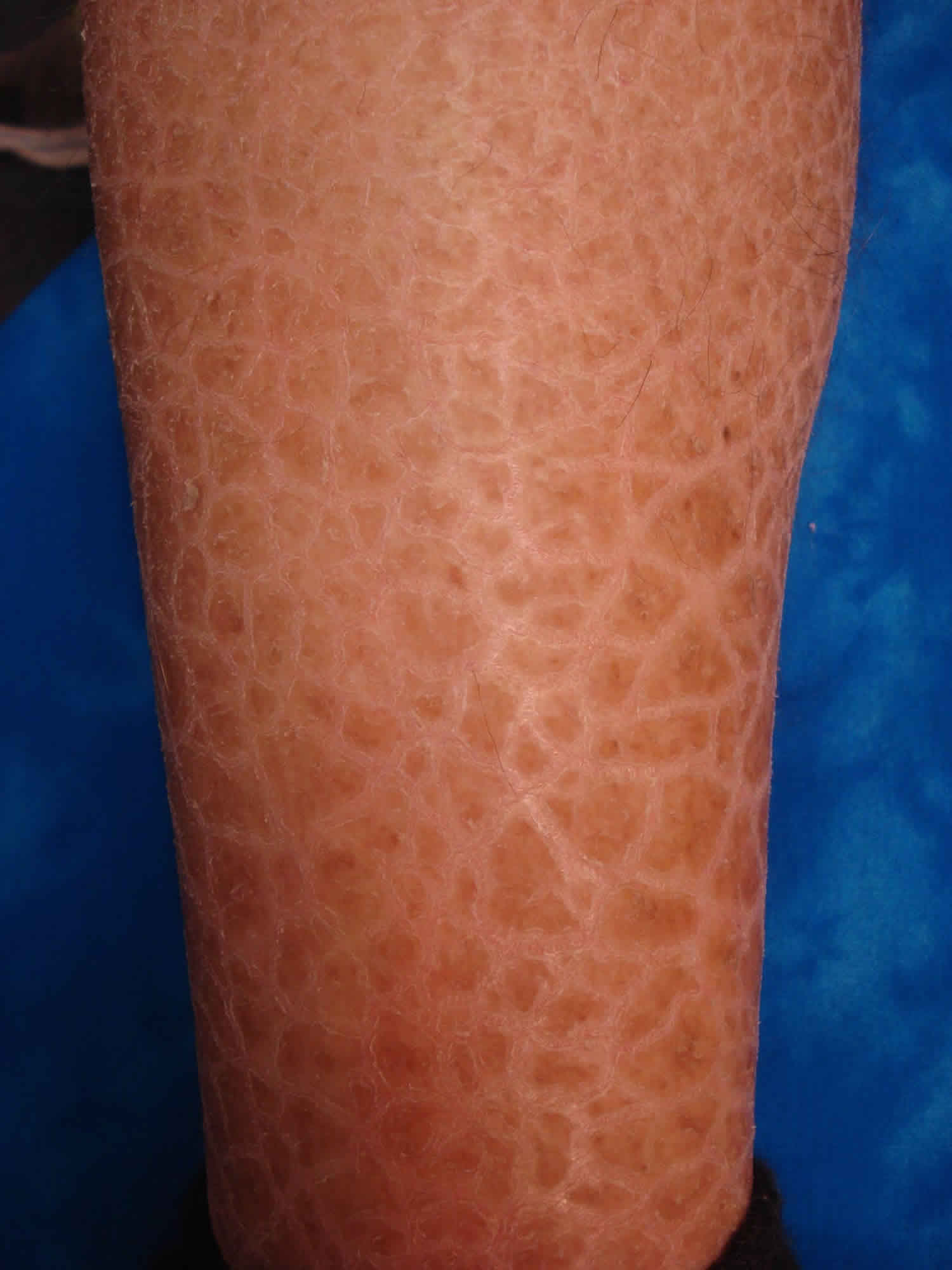
Harlequin ichthyosis is a severe, life-threatening genetic skin disorder characterized by hard, thick diamond-shaped plates of skin that crack and split, deeply affecting the body's primary protective barrier. While media profiles and individual stories—such as public accounts featuring individuals named Anna—bring personal visibility to rare conditions, understanding the underlying pathophysiology, inheritance patterns, and intensive multidisciplinary care requirements remains essential for navigating this complex medical reality.
Defining Harlequin Ichthyosis: Pathophysiology and Clinical Presentation
Harlequin ichthyosis is classified as the most severe form of congenital ichthyosis, a heterogeneous group of keratinization disorders. At the core of the condition is a profound disruption in the normal development of the stratum corneum, the outermost layer of the human epidermis.
Cellular Mechanisms and Lipid Transport
In healthy skin, epidermal cells continuously migrate upward from the basal layer, flattening and producing lipids that are packed into lamellar granules. These granules secrete lipids into the intercellular spaces, forming a waterproof, hydrophobic barrier that prevents excessive transepidermal water loss and blocks microbial invasion.
In individuals with harlequin ichthyosis, mutations occur within the ABCA12 gene (ATP-binding cassette subfamily A member 12). This gene provides instructions for producing a critical transporter protein responsible for moving lipids into lamellar granules in keratinocytes. When ABCA12 is defective or absent, these lipids cannot be properly transported. Consequently, the intercellular lipid lamellae fail to form, leading to a severely defective epidermal barrier and the hyper-accumulation of aberrant keratin layers.
Gross Physical Manifestations
At birth, infants with harlequin ichthyosis present a striking and distinct clinical appearance:
- Armor-Like Plates: The entire body is encased in thick, yellowish-white keratin plates separated by deep, red fissures or cracks.
- Craniofacial Distortion: The mechanical tightness of the skin plates causes severe ectropion (eversion of the eyelids), eclabion (eversion of the lips), and flattening of the external ears and nose. The eyelids often cannot close fully, placing the corneas at high risk of drying, ulceration, and infection.
- Compromised Thermoregulation and Hydration: The rigid encasement restricts normal chest expansion, leading to potential respiratory restriction. Furthermore, the skin barrier failure results in massive transepidermal water loss, severe electrolyte imbalances, and an inability to regulate core body temperature effectively.
Genetic Inheritance and Molecular Diagnostics
Harlequin ichthyosis follows an autosomal recessive inheritance pattern. This means an affected individual must inherit two mutated copies of the ABCA12 gene—one from each carrier parent.
Autosomal Recessive Mechanics
- Carrier Status: Parents of an affected child are typically asymptomatic carriers possessing one normal gene and one mutated gene.
- Recurrence Risk: With each pregnancy, carrier parents face a 25% chance of having a child with harlequin ichthyosis, a 50% chance of having an asymptomatic carrier child, and a 25% chance of having a child with two normal genes.
Diagnostic Verification
Diagnosis is typically suspected immediately at birth based on the distinct physical phenotype. Confirmation is achieved through:
- Genetic Testing: Molecular genetic testing via panel sequencing or targeted mutation analysis identifies pathogenic variants in the ABCA12 gene.
- Skin Biopsy: Histopathological examination of skin samples reveals compact hyperkeratosis and abnormal lamellar granule ultrastructure under electron microscopy, though genetic confirmation is increasingly standard.
- Prenatal Testing: For families with a known history or a previously affected child, prenatal diagnosis can be performed via chorionic villus sampling (CVS) or amniocentesis coupled with molecular genetic analysis.
Multidisciplinary Neonatal and Long-Term Management
Historically, harlequin ichthyosis carried an extremely high mortality rate in the neonatal period due to sepsis, dehydration, and respiratory failure. Modern advancements in neonatal intensive care units (NICUs) have significantly improved survival rates, shifting the focus toward aggressive, lifelong supportive management.
Acute Neonatal Care Protocols
The immediate priorities upon delivery in a specialized center involve stabilizing the infant and protecting fragile organ systems:
- Infection Prevention: Because the skin fissures create open pathways for pathogens, infants are typically managed in humidified incubators to minimize skin cracking and infection risks, accompanied by strict sterile nursing protocols.
- Ophthalmological Protection: Frequent application of lubricating eye drops and ointments, and occasionally temporary surgical closure of the eyelids (tarsorrhaphy), protects the exposed corneas.
- Nutritional Support: Feeding difficulties due to eclabion and restricted oral movement often require nasogastric or orogastric tube feeding to ensure adequate caloric intake and hydration.
- Respiratory Monitoring: Continuous monitoring of oxygen saturation and respiratory effort is vital, as the tightness of the chest wall can restrict lung expansion.
Systemic Retinoid Therapy
The introduction of oral systemic retinoids (such as acitretin) has revolutionized the management of harlequin ichthyosis. These medications help normalize keratinization, promoting the shedding of the thick hyperkeratotic plates. However, administration requires careful monitoring by pediatric dermatologists due to potential systemic side effects involving the liver, bones, and lipid metabolism.
| Management Phase | Primary Clinical Goals | Key Interventions & Monitoring |
|---|---|---|
| Neonatal Stabilization | Prevent infection, maintain fluid balance, protect corneas | Humidified incubators, sterile wound care, lubricating eye drops, tube feeding |
| Keratolytic Therapy | Soften and gently shed thick keratin scales | Topical emollients, keratolytics (e.g., urea, lactic acid), systemic retinoids |
| Long-Term Maintenance | Prevent skin fissuring, manage contractures, support development | Daily bathing and desquamation routines, physical therapy, nutritional optimization |
Daily Skincare, Maintenance, and Quality of Life
As children survive infancy, daily life requires a rigorous, time-intensive regimen focused on skin hydration and mechanical debridement.
The Daily Regimen
- Bathing and Soaking: Patients often require long, daily soaking baths to soften the hardened keratin plates. Gentle mechanical removal of loosened scale using soft cloths or sponges is performed carefully to avoid raw, bleeding skin.
- Emollient Application: Thick, preservative-free moisturizers, petroleum-based ointments, and barrier creams must be applied multiple times throughout the day to lock in moisture and mimic the missing lipid barrier.
- Environmental Controls: Maintaining stable ambient temperatures and humidity levels helps prevent overheating and excessive moisture loss, as individuals with ichthyosis often have impaired sweating capabilities due to duct occlusion by scales.
Psychosocial and Developmental Considerations
Living with a visible, rare dermatological condition presents notable social and psychological challenges. Comprehensive care involves:
- Dermatological Support: Access to specialized pediatric dermatology clinics experienced in barrier disorders.
- Psychological Counseling: Supporting patients and families through visible differences, social stigma, and the demands of chronic disease management.
- Physical Therapy: Addressing joint mobility and preventing contractures caused by tight skin over joints.
Frequently Asked Questions
What causes harlequin ichthyosis?
Harlequin ichthyosis is caused by mutations in the ABCA12 gene, which is essential for transporting lipids into cellular structures that form the skin's moisture and protective barrier. This genetic defect leads to abnormal thickening of the stratum corneum and the formation of rigid keratin plates.
Is harlequin ichthyosis contagious?
No, harlequin ichthyosis is a non-contagious, congenital genetic disorder. It is inherited in an autosomal recessive manner, meaning a child must inherit two mutated genes from carrier parents. It cannot be contracted from another person.
Can individuals with harlequin ichthyosis lead active lives?
Yes, with modern neonatal intensive care, aggressive skin hydration protocols, and systemic retinoid therapy, many individuals with harlequin ichthyosis survive infancy and grow up to attend school, pursue careers, and participate in daily activities, albeit with a lifelong requirement for intensive skincare routines.
How is harlequin ichthyosis diagnosed before birth?
Prenatal diagnosis can be performed when there is a known family history by using molecular genetic testing on samples obtained via chorionic villus sampling (CVS) or amniocentesis, allowing parents and medical teams to prepare for specialized delivery in a tertiary care center.
Are there any permanent cures for harlequin ichthyosis?
Currently, there is no permanent genetic cure for harlequin ichthyosis. Management focuses entirely on supportive care, barrier restoration, gentle exfoliation, and the prevention of secondary complications such as systemic infections and severe dehydration.
Conclusion and Next Actions
Navigating harlequin ichthyosis requires an immediate, coordinated medical response at birth followed by a lifelong commitment to intensive barrier maintenance and dermatological care. Families seeking guidance or facing a diagnosis should connect with specialized pediatric geneticists, dermatologists, and rare disease support organizations to access up-to-date treatment protocols and peer networks.