The primary difference between an alpha helix and a beta helix lies in their hydrogen bonding patterns and overall geometry. An alpha helix is a tight, single-stranded coil stabilized by internal hydrogen bonds between every fourth residue, while a beta helix is a larger, "solenoid" structure formed by multiple parallel beta strands winding around a central axis.
Fundamentals of Protein Secondary Structure
Protein architecture is organized into a hierarchy, beginning with the linear sequence of amino acids (primary structure) and progressing to local folding patterns known as secondary structures. These patterns are dictated by the rotational freedom of the polypeptide backbone, specifically the phi ($\phi$) and psi ($\psi$) dihedral angles. The most prevalent and well-studied of these patterns is the alpha helix. However, more complex repetitive motifs like the beta helix play critical roles in specialized biological functions, particularly in enzymes and structural proteins.
Understanding the distinction between an alpha helix and a beta helix requires looking beyond the word "helix." In biochemistry, a helix simply refers to a repeating spiral symmetry. While the alpha helix is a fundamental building block of almost all globular proteins, the beta helix is a specialized fold that behaves more like a rigid scaffold. In 2026, as computational protein design reaches new heights of precision, distinguishing these motifs is essential for synthetic biology and therapeutic development.
The Alpha Helix: The Ubiquitous Spiral
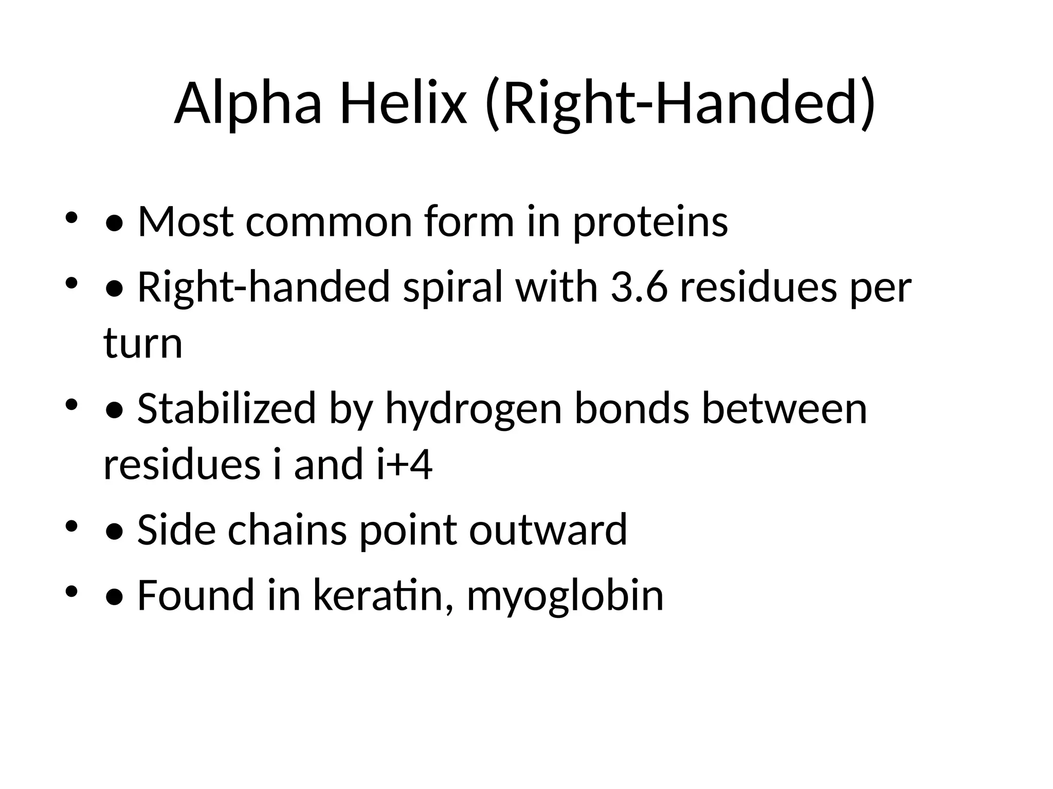
The alpha helix ($\alpha$-helix) is the most common secondary structure found in proteins. First proposed by Linus Pauling, Robert Corey, and Herman Branson in 1951, it is characterized by a right-handed coiled conformation in which every backbone N-H group donates a hydrogen bond to the backbone C=O group of the amino acid located four residues earlier ($i \rightarrow i+4$ hydrogen bonding).
Chemical Bonding and Stability
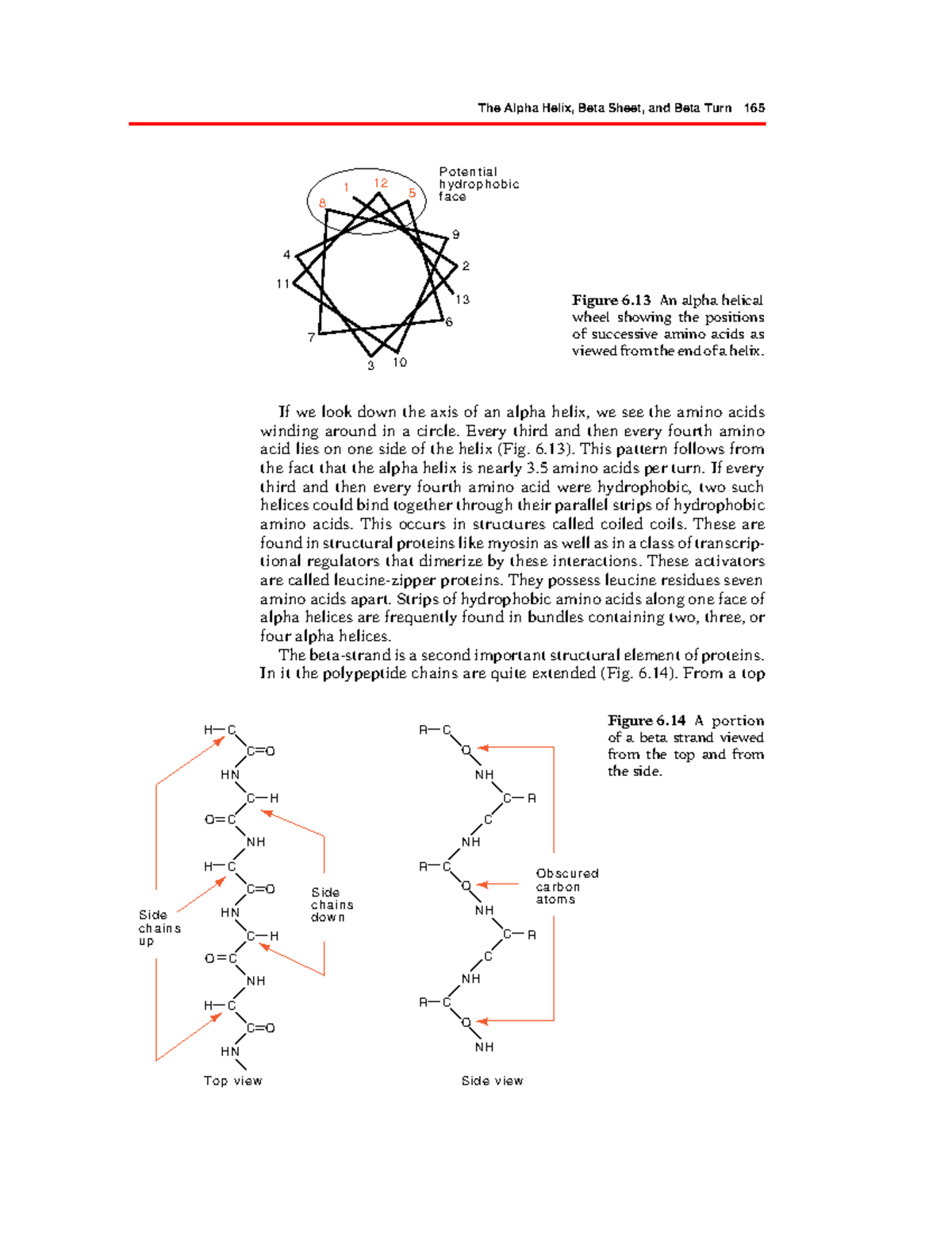
The stability of the alpha helix is derived from its repetitive hydrogen-bonding network. Because these bonds are parallel to the helical axis, they reinforce the structure like vertical struts in a tower. A standard alpha helix has 3.6 amino acid residues per turn, with a pitch (the vertical distance the helix rises per turn) of 5.4 Å. This arrangement ensures that the side chains (R-groups) of the amino acids point outward from the helical cylinder.
This outward orientation of side chains is crucial for the protein's interaction with its environment. For instance, in transmembrane proteins, alpha helices often consist of hydrophobic amino acids that interact with the lipid bilayer. In coiled-coil motifs, the outward-facing side chains of two different helices interlock like the teeth of a zipper, creating exceptionally strong fibrous structures such as keratin.
Amino Acid Propensities
Not all amino acids are equally likely to form an alpha helix. Alanine, leucine, and methionine are considered "helix formers" due to their favorable energetic profiles. Conversely, proline is often called a "helix breaker" because its cyclic side chain prevents the N-H group from participating in hydrogen bonding and introduces a physical "kink" in the backbone. Glycine, with its high conformational flexibility, also tends to destabilize the rigid alpha-helical structure unless specifically required for structural pivots.
The Beta Helix: A Robust Structural Motif
While the alpha helix is a single, continuous spiral of the polypeptide chain, the beta helix is a much larger and more complex arrangement. It belongs to the "solenoid" class of protein folds. A beta helix is formed by a single polypeptide chain that winds in a helical fashion, but the "rungs" of this helix are composed of short, parallel beta-strands connected by loops or turns.
Parallel Beta-Strands and Turns
In a beta helix, the polypeptide chain folds back on itself multiple times. This creates a structure that looks like a flattened or triangular prism. Unlike the alpha helix, where hydrogen bonds occur within a single segment of the coil, the beta helix is stabilized by hydrogen bonds between the beta-strands of adjacent "coils" or rungs.
Most beta helices are characterized by their "parallel" nature—meaning the N-to-C direction of the strands in each rung is the same. This creates a very stable, rigid, and elongated structure. The interior of the beta helix is often packed with hydrophobic side chains, creating a dry, stable core that can withstand harsh environments, such as the digestive tracts where many pectate lyases (common beta-helix enzymes) operate.
Right-handed vs Left-handed Beta Helices
Beta helices are classified based on their direction of winding.
- Right-handed Beta Helices: These are commonly found in enzymes like pectate lyases and certain viral tailspike proteins. They typically have a triangular cross-section.
- Left-handed Beta Helices: Often found in acyltransferases and carbonic anhydrases. These structures are typically more rounded or hexagonal in their cross-section compared to their right-handed counterparts.
The beta helix is notably larger in diameter than the alpha helix. While an alpha helix is a narrow rod, a beta helix is a hollow or solid-core cylinder that can serve as a scaffold for complex enzymatic active sites or as a mechanical drill in the case of certain bacteriophages.
Key Differences: Alpha Helix vs Beta Helix
The following table summarizes the fundamental structural and chemical differences between these two motifs as understood in contemporary structural biology.
| Feature | Alpha Helix | Beta Helix |
|---|---|---|
| Basic Unit | Single continuous coil | Multiple beta-strands and turns |
| Hydrogen Bonding | Intrastrand ($i \rightarrow i+4$) | Inter-rung (between adjacent strands) |
| Residues per Turn | 3.6 residues | Variable (often 15–30+ residues) |
| Diameter | Narrow (approx. 5 Å excluding side chains) | Wide (approx. 15–30 Å) |
| Side Chain Direction | Pointing outward from the axis | Pointing both inward (to core) and outward |
| Handedness | Almost exclusively right-handed | Can be right-handed or left-handed |
| Common Function | Signaling, DNA binding, transmembrane | Enzymatic catalysis, structural rigidity |
| Stability | High local stability | Exceptional global/mechanical stability |
Energetics and Thermodynamic Stability
The thermodynamic stability of these two structures is achieved through different means. The alpha helix relies on the cumulative strength of many weak hydrogen bonds aligned along the axis. Because it is a local structure, an alpha helix can fold almost instantaneously as the ribosome synthesizes the protein. This makes it a kinetically favored motif.
The beta helix, however, requires a more complex folding pathway. Since it involves interactions between segments of the chain that may be far apart in the primary sequence (though appearing in sequential rungs), the folding of a beta helix is often assisted by molecular chaperones. Once folded, the beta helix is incredibly robust. The "stacking" of beta-strands creates a repetitive, modular framework that is resistant to denaturation by heat or chemicals. This is why many extremophile bacteria utilize beta-helix motifs in their surface proteins and extracellular enzymes.
Biological Roles and Examples
The functional divergence between these structures is vast.
Alpha Helix Roles
- DNA Binding: The "recognition helix" in helix-turn-helix motifs fits perfectly into the major groove of DNA, allowing proteins to read genetic sequences.
- Transmembrane Anchors: Hydrophobic alpha helices are the standard mechanism for anchoring receptors and ion channels within the cell membrane.
- Mechanical Elasticity: In alpha-keratin (found in hair and wool), the stretching of alpha helices provides a degree of elasticity.
Beta Helix Roles
- Enzymatic Scaffolding: The rigid sides of the beta helix provide an ideal platform for arranging amino acids in a precise geometry to form active sites. This is seen in carbohydrate-active enzymes (CAZymes).
- Pathogenesis: Many bacterial toxins and viral attachment proteins use beta helices to maintain structural integrity while invading host cells. The "tailspike" of the P22 bacteriophage is a classic example of a beta-helix-rich protein that must survive harsh environmental transitions.
- Antifreeze Proteins: Some insects and fish produce antifreeze proteins that utilize a beta-helix structure to create a flat surface that binds to ice crystals, preventing them from growing and damaging tissues.
Computational Modeling and Prediction in 2026
As of 2026, the ability to predict and design these structures has reached near-experimental accuracy. While early versions of AI-driven folding models struggled with the repetitive nature of solenoid proteins like the beta helix, current iterative architectures can now distinguish between the subtle sequence variations that lead to a left-handed vs. a right-handed beta helix.
In de novo protein design, alpha helices are often used as "plug-and-play" modules for creating bundles. In contrast, beta helices are increasingly used as templates for designing "protein nanotubes" and "nanowires." Their hollow-core potential allows bioengineers to design proteins that can transport ions or small molecules through a controlled interior channel, mimicking natural porins but with the added stability of the beta-helix fold.
Frequently Asked Questions
Is a beta helix the same as a beta sheet?
No. A beta sheet is a secondary structure where strands are arranged side-by-side in a flat or slightly twisted plane. A beta helix is a specific 3D fold where a single chain winds around an axis to form a "solenoid" shape made of many short beta strands. You can think of a beta helix as a beta sheet that has been rolled into a coil.
Why is the alpha helix more common than the beta helix?
The alpha helix is a more "economical" fold. It requires only a short local sequence of amino acids to stabilize itself. The beta helix requires a much longer polypeptide chain and a specific repetitive sequence to form the rungs of the solenoid, making it a more specialized and rarer architectural choice in the proteome.
Can an amino acid be part of both an alpha helix and a beta helix?
Not simultaneously in the same protein molecule. An amino acid residue adopts specific $\phi$ and $\psi$ angles that define its secondary structure. However, a single protein can contain different domains where one domain is predominantly alpha-helical and another domain contains a beta helix.
What is the role of the central core in a beta helix?
The central core of a beta helix is usually packed with hydrophobic side chains (like isoleucine or valine) or sometimes organized "ladders" of polar residues. This packing is essential for the structure’s rigidity. In some engineered proteins, the core is modified to bind specific metal ions or small molecules.
How does handedness affect protein function?
In alpha helices, right-handedness is dictated by the L-amino acids that make up natural proteins; a left-handed alpha helix is energetically unfavorable. In beta helices, both right-handed and left-handed versions exist because the "handedness" refers to the overall twist of the rungs, which can be accommodated by the peptide backbone in both directions depending on the turn sequences.
Practical Recommendations for Structural Analysis
When analyzing a new protein sequence for these motifs, researchers should look for specific patterns:
- Periodicity: Alpha helices show a strong 3.6-residue periodicity in hydrophobicity if they are amphipathic. Beta helices often exhibit a longer, more complex repeating sequence (often involving hexapeptide or octapeptide repeats).
- Circular Dichroism (CD) Spectroscopy: Alpha helices provide a distinct "double-dip" signature at 208 nm and 222 nm. Beta helices, being composed of beta strands, produce a signature more similar to beta sheets (a single minimum around 218 nm), though the "solenoid" nature can sometimes shift these values.
- Structural Context: If the protein is a known extracellular enzyme (like a lipase or lyase) or a viral structural protein, the probability of encountering a beta-helix motif increases significantly compared to intracellular signaling proteins.
Understanding these differences is not merely an academic exercise; it is the foundation for interpreting how proteins function as molecular machines and how we can redesign them for the future of biotechnology.