Afferent lymphatic vessels are specialized vascular structures that transport lymph fluid from peripheral tissues and interstitial spaces into the convex surface of a lymph node. These vessels serve as the primary entry points for antigens, pathogens, and immune cells, allowing the lymph node to filter fluid and initiate targeted immune responses.
The Anatomical Framework of Afferent Lymphatic Vessels
The lymphatic system is a complex network of vessels, tissues, and organs that maintains fluid homeostasis and supports the immune system. At the heart of this network is the afferent lymphatic vessel. The term "afferent" is derived from the Latin afferens, meaning "carrying toward." In the context of the lymphatic system, these vessels carry unfiltered lymph toward the lymph nodes, which act as biological filtration stations.
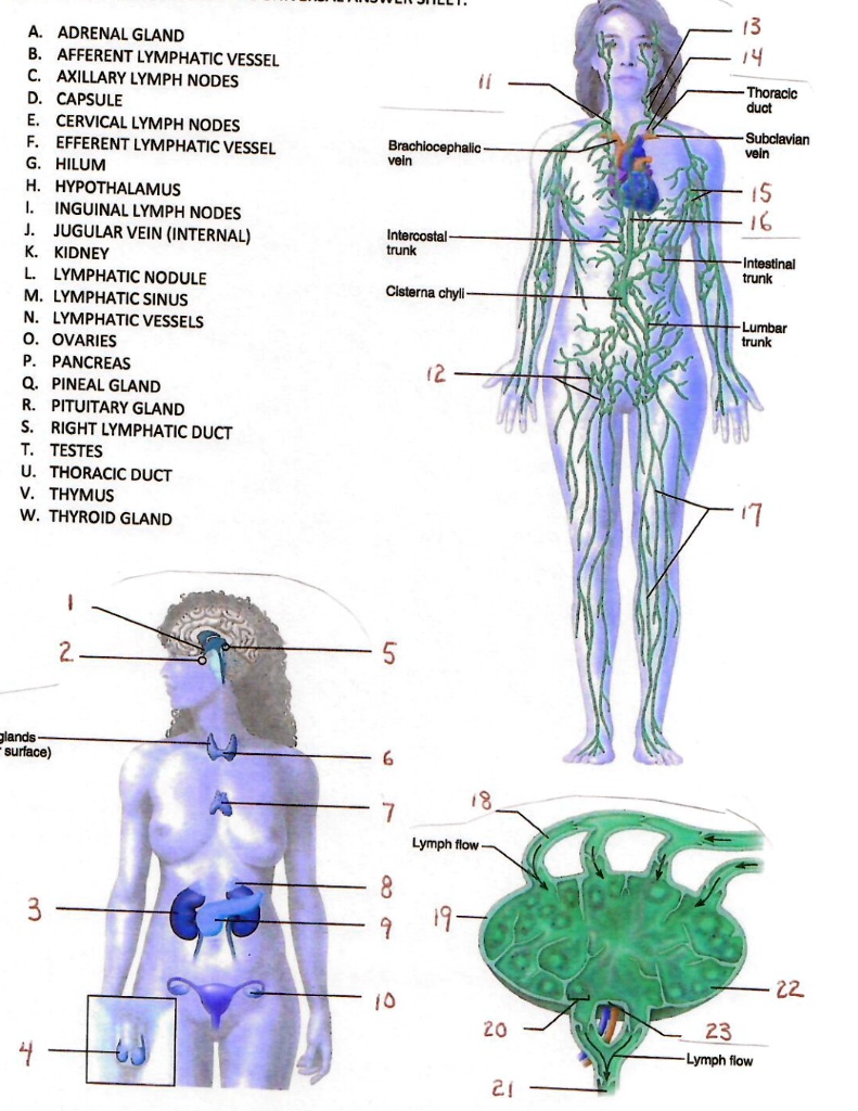
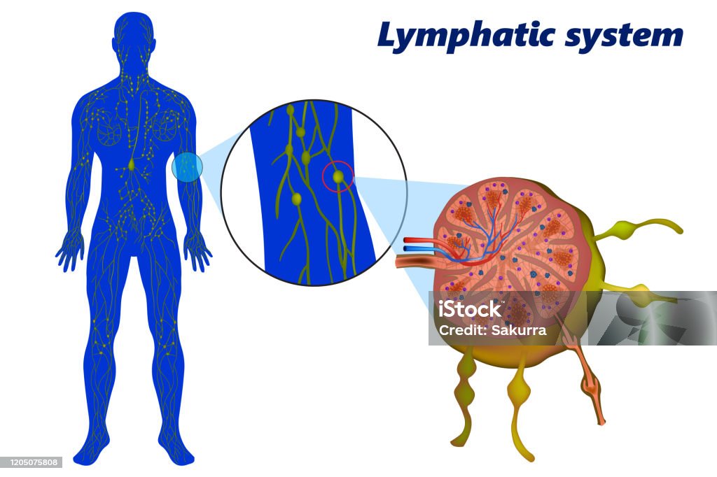
Anatomically, afferent vessels enter the lymph node at multiple points along its outer, convex capsule. This is a distinct structural characteristic; while several afferent vessels bring fluid into a single node, typically only one or two efferent vessels carry the filtered lymph out through the hilum (the indented region of the node). This numerical disparity is functionally significant, as it slows the flow of lymph within the node, providing ample time for macrophages and lymphocytes to interact with any foreign particles or pathogens present in the fluid.
The walls of afferent lymphatic vessels are remarkably thin, composed primarily of a single layer of endothelial cells. Unlike the high-pressure environment of the arterial system, the lymphatic system operates under low pressure. To manage this, afferent vessels contain one-way valves. These semilunar valves ensure that once lymph enters the vessel, it can only move toward the lymph node, preventing retrograde flow even when body positioning or external pressure changes.
The Journey of Lymph: From Interstitial Space to the Node
The process begins in the peripheral tissues, where interstitial fluid—fluid that has leaked out of blood capillaries—collects in the spaces between cells. Once this fluid enters the initial lymphatic capillaries, it is officially termed lymph. As these capillaries merge, they form larger collecting vessels, which eventually become the afferent lymphatic vessels.
The composition of lymph entering an afferent vessel is a reflection of the tissue it just drained. It contains water, electrolytes, proteins, and lipids, but it also carries "debris" such as cellular waste, bacteria, viruses, and even malignant cells if a tumor is present. Most importantly, afferent vessels transport dendritic cells—professional antigen-presenting cells that have "captured" foreign proteins in the tissue and are now traveling to the lymph node to "present" them to T-cells and B-cells.
The entry of lymph into the node via afferent vessels is not a simple dump of fluid. The vessels penetrate the capsule and empty into the subcapsular sinus. From there, the fluid trickles through a mesh-work of connective tissue fibers (reticular fibers) and pockets of immune cells in the cortex and medulla. This slow percolation is essential for the "surveillance" function of the immune system.
Physiological Mechanisms Driving Lymphatic Flow
Because the lymphatic system lacks a central pump like the heart, the movement of lymph through afferent vessels relies on several extrinsic and intrinsic mechanisms. As of 2026, clinical understanding emphasizes the importance of the "lymphatic pump" or lymphangion—the segment of a lymphatic vessel between two valves.
- Intrinsic Contraction: The smooth muscle cells within the walls of larger afferent vessels undergo spontaneous, rhythmic contractions. When a lymphangion fills with fluid, the stretching of the vessel wall triggers a contraction that pushes the fluid through the next valve.
- Skeletal Muscle Pump: Movement of the limbs compresses the thin-walled afferent vessels. This external pressure squeezes the lymph forward. This is why physical activity is often recommended to reduce minor swelling and improve immune circulation.
- Respiratory Pump: During inhalation, the pressure in the thoracic cavity decreases while the pressure in the abdominal cavity increases. This pressure gradient helps "pull" lymph upward toward the lymphatic ducts.
- Arterial Pulsation: Many afferent lymphatic vessels travel alongside deep arteries. The rhythmic pulsing of the arteries provides a subtle but constant external pressure that aids in moving lymph toward the nodes.
Functional Significance in Immune Activation
The primary role of the afferent lymphatic vessel bringing lymph into a lymph node is to facilitate the detection of threats. The lymph node is essentially a specialized environment for cellular communication. When afferent vessels deliver antigens to the node, the following sequence occurs:
- Filtration: Macrophages residing in the lymph node sinuses engulf and digest bacteria, viruses, and debris delivered by the afferent stream.
- Antigen Presentation: Dendritic cells that traveled via the afferent vessels migrate into the T-cell zones of the node. They display fragments of the pathogens they found in the periphery to "naive" T-lymphocytes.
- Clonal Expansion: If a T-cell recognizes the antigen, it becomes activated and begins to divide rapidly, preparing the body for a systemic immune response.
- B-cell Activation: Antigens also reach the follicles where B-cells reside. This leads to the production of antibodies, which will eventually leave the node via the efferent vessel to circulate in the bloodstream.
Without the afferent vessels acting as the "inbound lane," the immune system would remain unaware of localized infections until they became systemic and life-threatening.
Structural and Functional Comparisons
To understand the unique role of the afferent vessel, it is helpful to compare it to the efferent vessel. While they share the same basic goal of fluid transport, their specific roles in the node's architecture are polarized.
| Feature | Afferent Lymphatic Vessel | Efferent Lymphatic Vessel |
|---|---|---|
| Direction of Flow | Toward the lymph node | Away from the lymph node |
| Point of Entry/Exit | Convex surface of the node | Concave surface (Hilum) |
| Quantity | Multiple vessels per node | Usually one or two vessels per node |
| Fluid Composition | Contains unfiltered debris and pathogens | Rich in activated lymphocytes and antibodies |
| Primary Function | Delivery of antigens for surveillance | Distribution of immune cells and filtered fluid |
| Valve Presence | Yes (to prevent backflow to tissues) | Yes (to prevent backflow to the node) |
Clinical Implications: When Afferent Transport Fails
The health and patency of afferent lymphatic vessels are critical. When these vessels are damaged or blocked, the results are often clinically significant.
Lymphedema
If afferent vessels are removed (as often happens during cancer surgery) or damaged by radiation, lymph fluid cannot reach the nodes. This leads to an accumulation of protein-rich fluid in the interstitial spaces, a condition known as lymphedema. Because the fluid is not being filtered by the nodes, the affected area also becomes more susceptible to infections like cellulitis.
Cancer Metastasis
The afferent lymphatic pathway is frequently hijacked by malignant cells. In many types of cancer, such as breast cancer or melanoma, tumor cells break away from the primary site and enter the afferent lymphatic capillaries. They are then carried to the "sentinel" lymph node—the first node in the drainage chain. Surgeons often perform sentinel lymph node biopsies to determine if cancer has begun to spread, as the afferent vessels are the most common route for early metastasis.
Lymphangitis
Inflammation of the afferent lymphatic vessels, often due to a bacterial infection (typically Streptococcus pyogenes), is known as lymphangitis. This often presents as visible red streaks on the skin extending from the site of an infection toward the nearest group of lymph nodes. It indicates that the afferent vessels are actively transporting pathogens and are becoming inflamed in the process.
Modern Research and Perspectives in 2026
By 2026, advances in high-resolution imaging and molecular biology have allowed scientists to observe the "trafficking" within afferent vessels in real-time. We now know that the endothelial cells of afferent vessels are not passive barriers; they actively secrete chemokines, such as CCL21, which act as chemical breadcrumbs for dendritic cells to follow into the vessel.
Furthermore, research into the "glymphatic system"—the waste-clearance system of the brain—has revealed how metabolic waste from the central nervous system eventually finds its way into the cervical afferent lymphatic vessels. This discovery has bridged the gap between neurology and immunology, highlighting the afferent vessel's role in maintaining cognitive health by facilitating the removal of amyloid-beta and tau proteins.
FAQ: Understanding Afferent Lymphatic Function
Why are there more afferent vessels than efferent vessels? This anatomical design is intentional. By having multiple "inbound" afferent vessels and only one or two "outbound" efferent vessels, the lymph node creates a bottleneck. This slows down the flow of lymph, ensuring the fluid is thoroughly filtered and that immune cells have enough time to interact with any antigens present.
Can lymph bypass the afferent vessels? In a healthy state, lymph must travel through the afferent vessels to reach a lymph node. However, in cases of severe blockage or surgical bypass (lymphatic-venous anastomosis), lymph may be diverted directly into the venous system. This is a compensatory or surgical intervention and is not the standard physiological route.
Do afferent vessels carry blood? No, afferent lymphatic vessels only carry lymph. While they often run parallel to blood vessels and share some structural similarities with veins (such as valves and low pressure), the lymphatic and circulatory systems are distinct. Lymph is typically clear or milky, whereas blood is red due to the presence of erythrocytes, which are generally absent in healthy lymph.
How do doctors check the health of afferent vessels? The most common method is lymphoscintigraphy, where a radioactive tracer is injected into the skin. Imaging then tracks the tracer as it moves through the afferent vessels to the lymph nodes. Other methods include Indocyanine Green (ICG) fluorescence imaging, which provides a real-time view of lymphatic flow.
Next Steps for Health Maintenance
Understanding the role of afferent lymphatic vessels highlights the importance of supporting lymphatic circulation. To maintain healthy lymphatic transport:
- Stay Hydrated: Lymph is mostly water; dehydration can cause lymph to become sluggish.
- Maintain Movement: Regular physical activity acts as a manual pump for afferent vessels.
- Monitor for Swelling: Any persistent swelling in the limbs should be evaluated by a healthcare professional to ensure afferent lymphatic drainage is not compromised.
- Skin Care: Since afferent vessels drain the skin, preventing and promptly treating small cuts or infections reduces the "load" of pathogens those vessels must transport to your nodes.
Note: This article is for informational purposes and does not constitute medical advice. For concerns regarding lymphatic health, consult a qualified healthcare provider.