An abdominal examination for suspected appendicitis focuses on identifying specific patterns of tenderness and localized peritonitis. Through systematic inspection, auscultation, percussion, and palpation—specifically targeting the right lower quadrant (RLQ)—clinicians evaluate for involuntary guarding, rebound tenderness, and classic physical signs like McBurney’s point tenderness to determine the necessity of urgent surgical consultation or diagnostic imaging.
Understanding the Clinical Presentation of Appendicitis
Acute appendicitis remains one of the most common surgical emergencies globally. The classic presentation—often starting as periumbilical pain that migrates to the right lower quadrant—is frequently accompanied by anorexia, nausea, and low-grade fever. However, clinicians must approach the abdominal examination with the understanding that the appendix is anatomically variable; it may be retrocecal, pelvic, or subhepatic, which can significantly alter the clinical findings and the results of a physical exam.
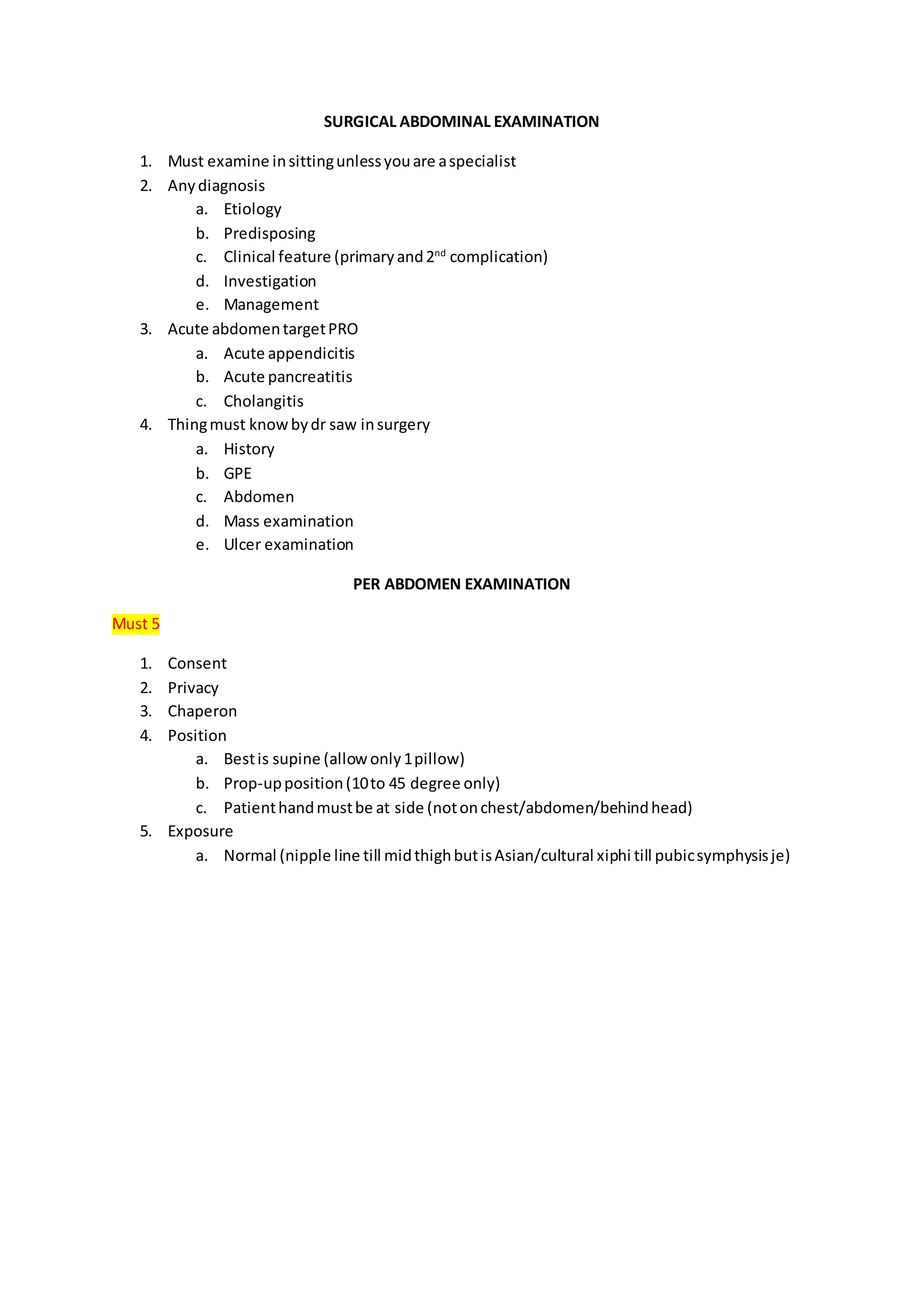
Before beginning the physical examination, the clinician must ensure the patient is positioned comfortably in the supine position with the abdomen exposed. A systematic approach is essential to differentiate appendicitis from other pathologies such as ovarian torsion, ectopic pregnancy, nephrolithiasis, or inflammatory bowel disease.
The Systematic Physical Examination Protocol
A formal examination of the abdomen follows a standard sequence: inspection, auscultation, percussion, and palpation. Each phase provides critical data that, when synthesized, informs the likelihood of an acute surgical abdomen.
1. Inspection
The examination begins with observing the patient’s overall demeanor. Patients with peritonitis often lie still, as movement—including coughing or bumpy car rides—exacerbates their pain. Inspect the abdomen for distention, visible surgical scars, or asymmetry. The patient should be asked to point to the area of maximal pain with a single finger; this "point tenderness" is a highly reliable indicator of localized peritoneal irritation.
2. Auscultation
While auscultation is less specific for appendicitis, it is vital for ruling out other causes of abdominal pain, such as mechanical bowel obstruction (indicated by high-pitched, hyperactive bowel sounds) or adynamic ileus (indicated by absent bowel sounds). In the case of early appendicitis, bowel sounds may remain normal or slightly diminished due to an associated localized ileus.
3. Percussion
Gentle percussion of the abdomen helps identify areas of localized peritonitis. The clinician should start away from the area of maximal pain and work toward it. If the patient experiences pain when the bed is gently tapped or when they are asked to cough (the "cough test"), this suggests peritoneal irritation. Percussion can also identify dullness, which may indicate an underlying fluid collection or an enlarged organ.
4. Palpation
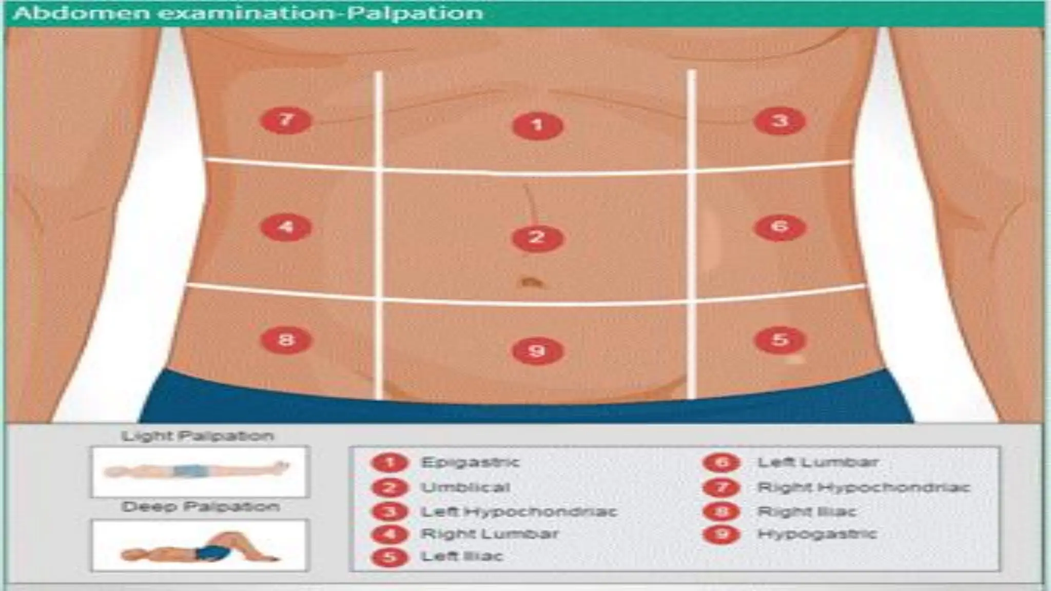
Palpation is the most diagnostic component of the exam. The clinician must use a gentle, warm hand to avoid triggering voluntary guarding, which can mask underlying findings.
- Superficial Palpation: Used to identify areas of general tenderness and muscle resistance.
- Deep Palpation: Performed to assess for masses, such as an appendiceal phlegmon or abscess.
- Rebound Tenderness (Blumberg’s Sign): This is elicited by applying steady pressure over the area of tenderness and then rapidly releasing it. A sudden increase in pain upon release suggests localized peritoneal inflammation.
Specialized Physical Signs for Appendicitis
Over the decades, several clinical maneuvers have been standardized to aid in the diagnosis of appendicitis. While no single sign is 100% sensitive or specific, the presence of these findings significantly increases the clinical suspicion.
| Sign | Execution | Interpretation |
|---|---|---|
| McBurney’s Point | Palpation 1/3 of the distance from the ASIS to the umbilicus. | Maximal tenderness here is a classic indicator of appendicitis. |
| Rovsing’s Sign | Pressure applied to the left lower quadrant. | Pain felt in the right lower quadrant indicates peritoneal irritation. |
| Psoas Sign | Passive extension of the patient’s right thigh while supine. | Pain indicates an inflamed retrocecal appendix irritating the psoas muscle. |
| Obturator Sign | Passive internal rotation of the flexed right hip. | Pain indicates an inflamed appendix in contact with the obturator internus. |
Challenges in Diagnosis: Anatomical Variability
The primary difficulty in performing an abdominal examination for appendicitis is the varying anatomical position of the appendix. When the appendix is located behind the cecum (retrocecal), the patient may present with flank pain rather than the classic RLQ pain, leading to potential confusion with renal colic. If the appendix is pelvic in orientation, the patient may present with diarrhea or dysuria rather than acute abdominal pain, as the inflammation irritates the bladder or rectum.
Furthermore, extremes of age present unique diagnostic hurdles. In pediatric patients, the omentum is shorter and less able to "wall off" an infection, leading to a higher risk of rapid progression to perforation and generalized peritonitis. In elderly patients, the physical exam findings are often muted, and symptoms may be atypical, frequently resulting in a delayed diagnosis and increased morbidity.
Integrating Diagnostic Imaging
In 2026, the physical examination remains the foundation of diagnosis, but it is almost universally supported by imaging in cases of diagnostic uncertainty. Clinical decision rules, such as the Alvarado Score or the Appendicitis Inflammatory Response (AIR) score, are often used to stratify patients into low, moderate, and high-risk groups.
If the abdominal examination findings are equivocal, imaging is required:
- Computed Tomography (CT): The gold standard for adults, providing high sensitivity and specificity for identifying inflammation, diameter of the appendix, and presence of perforation or abscess.
- Ultrasound: The preferred modality for pediatric patients and pregnant women to avoid ionizing radiation. It is highly operator-dependent but effective when the appendix is clearly visualized.
- Magnetic Resonance Imaging (MRI): Increasingly utilized for pregnant patients when ultrasound is inconclusive, as it provides excellent soft-tissue contrast without radiation risk.
Common Mistakes and How to Avoid Them
- Failing to Re-examine: Appendicitis is a dynamic process. If the diagnosis remains unclear, serial physical examinations conducted by the same clinician every few hours are critical to track the evolution of symptoms.
- Over-relying on "Classic" Symptoms: Never rule out appendicitis simply because the patient lacks a fever or presents with atypical pain. The appendix is a fickle organ; assume the worst until proven otherwise.
- Ignoring the "Pain before Vomiting" Sequence: In classic appendicitis, the onset of abdominal pain typically precedes nausea and vomiting. If vomiting occurs before the onset of pain, consider other causes like gastroenteritis.
- Inadequate Exposure: A proper abdominal examination cannot be performed through clothing. Ensure the patient is properly draped and the entire abdomen is visible to allow for accurate palpation.
FAQ: Clinical Considerations
How reliable is McBurney’s point tenderness in diagnosing appendicitis? While McBurney’s point is the classic site of tenderness, it is not diagnostic in isolation. Its presence should be interpreted in the context of the entire physical exam, including other signs like the psoas or obturator signs, and the patient's overall clinical history.
Can an abdominal exam rule out appendicitis entirely? No. A physical examination cannot rule out appendicitis with 100% certainty, particularly in early stages or in patients with retrocecal or pelvic appendices. If the clinical index of suspicion remains high despite a "negative" exam, further observation or diagnostic imaging is mandated.
What is the role of the Alvarado Score in clinical practice? The Alvarado Score is a clinical scoring system that combines symptoms (migratory pain, anorexia), signs (tenderness, rebound, fever), and lab data (leukocytosis, shift to left). It helps standardize the decision-making process, helping clinicians distinguish between patients who require immediate surgery and those who require further observation.
Why is pain often worse when coughing during appendicitis? Coughing causes a sudden increase in intra-abdominal pressure and movement of the peritoneum. If the parietal peritoneum is inflamed due to an underlying appendicitis, this movement results in a sharp, localized, and involuntary pain response.
Should I provide pain medication during an appendectomy evaluation? Modern evidence suggests that providing appropriate analgesia does not mask the findings of an abdominal examination or delay the diagnosis of surgical conditions. Withholding pain management can cause unnecessary distress and may actually make a physical exam more difficult if the patient is unable to cooperate due to severe pain.
Next Steps and Recommendations
If you are evaluating a patient for potential appendicitis, prioritize a structured physical assessment. Document the presence or absence of each specific maneuver, such as the psoas or Rovsing's sign, in the clinical record. If you are a patient experiencing sharp, persistent abdominal pain that migrates to the lower right side, seek immediate evaluation at an emergency department. Do not consume food or water, as you may require urgent surgical intervention. In cases where the clinical picture is unclear, expect the medical team to utilize serial exams, laboratory blood work, and appropriate imaging to ensure an accurate and timely diagnosis.