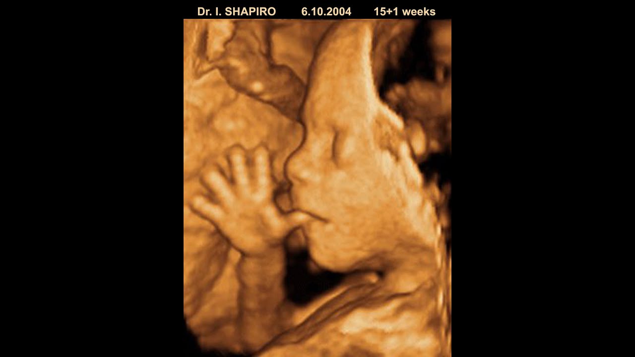
At 20 weeks gestation, a 3D ultrasound provides a surface-rendered, life-like perspective of the fetus, offering a significant visual upgrade from the traditional two-dimensional grayscale views. While 2D imaging remains the diagnostic gold standard for evaluating internal organ health and structural development during the anatomy scan, 3D technology enhances the ability to visualize the fetus's facial features, skin surface, and limbs.
Understanding the Difference Between 2D, 3D, and 4D Ultrasound
To appreciate what you are looking at during a 20-week scan, it is essential to distinguish between the various imaging modes. Most clinical anatomy scans are conducted primarily in 2D.
Traditional 2D Ultrasound
This is the standard mode used by technicians and radiologists. It produces a flat, cross-sectional, grayscale image. Because it captures a single plane at a time, it is highly effective at showing internal structures like the heart chambers, kidneys, brain ventricles, and the spine. Clinicians rely on 2D images to measure the growth of the fetus and ensure that developmental milestones are met.
3D Ultrasound Imaging
3D technology works by acquiring multiple 2D images from different angles and processing them through a computer to create a volumetric reconstruction. This results in a static, three-dimensional image that appears more like a photograph. At 20 weeks, you can often see the contours of the nose, the shape of the lips, and the position of the ears.
4D Ultrasound
4D imaging is essentially 3D ultrasound in real-time. It adds the dimension of time, allowing you to see the fetus moving, yawning, or sucking its thumb. While this is popular for "keepsake" scans, it relies on the same acoustic technology as 3D imaging.
Why 20 Weeks is a Strategic Time for Imaging
The 20-week mark—often referred to as the "mid-pregnancy" or "anatomy scan"—is the ideal window for viewing these images. By this stage, the fetus has developed distinct facial features and, importantly, there is still enough amniotic fluid surrounding the fetus to act as a window for the sound waves.
Adequate Amniotic Fluid
Sound waves used in ultrasound travel poorly through bone or dense tissue but move efficiently through fluid. At 20 weeks, the ratio of amniotic fluid to the size of the fetus is optimal. As the pregnancy progresses toward the third trimester, the fetus occupies more space in the uterus, often pressing its face against the uterine wall or the placenta, which can obstruct the view and create shadows.
Fetal Development
By 20 weeks, the fetus has accumulated enough subcutaneous fat to define the contours of the face. While the fetus will continue to fill out, the 20-week scan provides a clear look at the structural formation that is already complete.
Factors Influencing Image Quality
It is important to manage expectations regarding the quality of 3D ultrasound images. Several variables can prevent the capture of a "perfect" photograph, and these are often beyond the control of the ultrasound technician.
Fetal Position
The position of the fetus is the single greatest variable. If the fetus is facing toward the mother’s spine, or if its hands, feet, or umbilical cord are positioned in front of the face, a clear 3D rendering will be difficult to achieve.
Maternal Body Habitus
The thickness of the abdominal wall and the amount of adipose tissue can affect the clarity of the sound waves. Because ultrasound must penetrate through the mother’s skin, muscle, and fat, a higher density of these tissues can cause signal attenuation, resulting in grainier images.
Amniotic Fluid Volume and Clarity
As mentioned, fluid is necessary for a good image. If there is low amniotic fluid (oligohydramnios), the sound waves will not have a clear path to reconstruct the 3D surface. Additionally, if the placenta is located on the front wall of the uterus (anterior placenta), it may block the view of the fetal face.
Comparison of Diagnostic vs. Keepsake Imaging
It is crucial to distinguish between a clinical anatomy scan and a commercial "keepsake" or "elective" ultrasound service.
| Feature | Diagnostic Scan (20 Weeks) | Elective/Keepsake Scan |
|---|---|---|
| Purpose | Medical assessment of fetal health | Entertainment/bonding experience |
| Personnel | Registered Diagnostic Medical Sonographer | Often non-medical technicians |
| Regulation | FDA approved standards for medical use | Limited or no medical oversight |
| Data Usage | Used for prenatal care decisions | Used for family keepsakes |
| Timing | Scheduled for medical necessity | Scheduled at owner's convenience |
The American College of Obstetricians and Gynecologists (ACOG) and the FDA advise that ultrasounds should only be performed when there is a clear medical justification. While there is no known evidence that ultrasound causes harm to the fetus, the principle of ALARA (As Low As Reasonably Achievable) should always be followed regarding exposure to acoustic energy.
What to Look for in Your 3D Images
When you look at a 3D ultrasound from your 20-week scan, you may initially be confused by the appearance. Unlike a standard photograph, a 3D ultrasound is a computer-generated reconstruction.
- Facial Structures: Look for the bridge of the nose and the cupids-bow shape of the lips. These are the most common landmarks visible at this stage.
- Volumetric Rendering: You may see "artifacts," which are visual errors where the computer has misinterpreted a shadow or a piece of umbilical cord as part of the fetal surface. Do not mistake these for physical abnormalities.
- Surface Shading: The computer applies light and shadow to the reconstruction to give it depth. If the "light" seems to come from an unnatural angle, it is simply the software’s way of highlighting the surface contours.
Managing Expectations and Limitations
Many parents look forward to the 20-week scan as a way to "see" their baby. However, it is essential to approach this with a balanced mindset.
Technical Limitations
3D ultrasound is not a medical diagnostic tool for every condition. It cannot see internal organs as clearly as 2D, and it is prone to visual distortion. If your clinician does not use 3D mode, it does not mean they are missing something; they are likely prioritizing the diagnostic accuracy of the 2D sweep.
Emotional Preparedness
If the fetus is in a difficult position, the technician may spend time trying to move the baby by having the mother change positions. Despite these efforts, a clear face shot is never guaranteed. Being prepared for a "non-photo-ready" session can prevent disappointment.
Frequently Asked Questions
Is 3D ultrasound safe for my baby?
Yes, when performed by a trained professional for medical purposes. The ultrasound technology used for 3D is the same as 2D; it simply uses advanced software to render the data. The FDA advises against using ultrasound for non-medical purposes, such as keeping a video, because it results in unnecessary exposure to sound waves.
Can 3D ultrasound detect birth defects?
While 3D imaging can sometimes provide a better visual of surface structures like cleft lips or limb anomalies, the primary detection of birth defects is done during the 2D anatomy scan. Doctors use 2D imaging to look at the heart, brain, and spine, which are the most critical areas for diagnosing potential issues.
Why does my baby look "alien" in the 3D scan?
The 3D ultrasound captures the fetus as it exists in the womb, often surrounded by fluid and lacking the full layer of baby fat that develops closer to the third trimester. Because the software is rendering a surface, skin folds or thin tissue can sometimes appear exaggerated, leading to a look that doesn't necessarily reflect how the baby will look at birth.
What should I do if my doctor says the images are unclear?
If a doctor or technician reports that the images are unclear, it is almost always due to the position of the baby, the location of the placenta, or maternal body habitus. It rarely indicates a problem with the fetus. If the anatomy scan is diagnostic, your doctor will rely on the 2D images to confirm health, making the 3D quality secondary.
Can I request a 3D scan if my hospital doesn't offer one?
Most hospitals prioritize diagnostic imaging. If your clinic does not perform 3D as part of their routine care, it is usually because they have determined it does not provide additional clinical value for your specific check-up. You should discuss your desire for 3D imaging with your OB-GYN before seeking an outside provider, as they may have concerns about the facility's safety standards.
Next Steps for Your Anatomy Scan
As you prepare for your 20-week appointment, remember that the primary goal is the thorough assessment of your fetus’s development.
- Confirm the appointment: Ensure you are well-hydrated, as adequate hydration can sometimes help improve the clarity of the amniotic fluid.
- Discuss preferences: Ask your provider in advance if they intend to use 3D imaging. If they do not, respect their medical judgment, as their priority is the health of the pregnancy.
- Focus on the report: After the scan, your provider will review the findings. The written report from the radiologist or your OB-GYN is the authoritative source of information regarding the status of your pregnancy.
- Limit elective imaging: If you choose to seek extra images through an elective service, ensure the facility is reputable and adheres to strict safety guidelines regarding the duration of the scan.