An air-fluid level behind the tympanic membrane is a clinical sign indicating that the middle ear space is partially filled with liquid—such as serum, mucus, or blood—while the remaining space contains air. This phenomenon, typically visualized during otoscopy, confirms middle ear effusion and often results from Eustachian tube dysfunction or barotrauma.
The visualization of an air-fluid level is a pivotal moment in an otological examination. It serves as a definitive diagnostic marker for middle ear effusion (MEE), distinguishing it from other ear pathologies that might present with similar symptoms like ear fullness or hearing loss but lack the physical presence of fluid. In the clinical environment of 2026, the identification of this level remains a fundamental skill for primary care physicians, pediatricians, and otolaryngologists alike.
Understanding the mechanics of why fluid accumulates, how it presents under various lighting conditions, and what it implies for a patient’s hearing and health is essential for determining the correct course of treatment. This guide provides an exhaustive analysis of the air-fluid level within the tympanic membrane, covering its pathophysiology, diagnostic techniques, and management strategies.
Anatomy and Physiology of the Middle Ear Space
To understand the formation of an air-fluid level, one must first understand the delicate balance of the middle ear. The middle ear is an air-filled cavity located within the temporal bone. It is separated from the external environment by the tympanic membrane (eardrum) and is connected to the nasopharynx by the Eustachian tube.
The Role of the Eustachian Tube
The Eustachian tube is the primary regulator of the middle ear environment. It performs three critical functions:
- Pressure Equalization: It opens intermittently to match the air pressure in the middle ear with the atmospheric pressure.
- Protection: It prevents the reflux of nasopharyngeal secretions and sound energy into the middle ear.
- Clearance/Drainage: It allows for the drainage of naturally occurring secretions from the middle ear lining into the nasopharynx via mucociliary transport.
When the Eustachian tube fails—due to inflammation, anatomical abnormalities, or rapid external pressure changes—a vacuum (negative pressure) is created within the middle ear. This negative pressure can cause the mucous membrane lining the middle ear to secrete fluid or draw serous fluid from surrounding tissues, leading to the accumulation that forms an air-fluid level.
Characteristics of the Tympanic Membrane
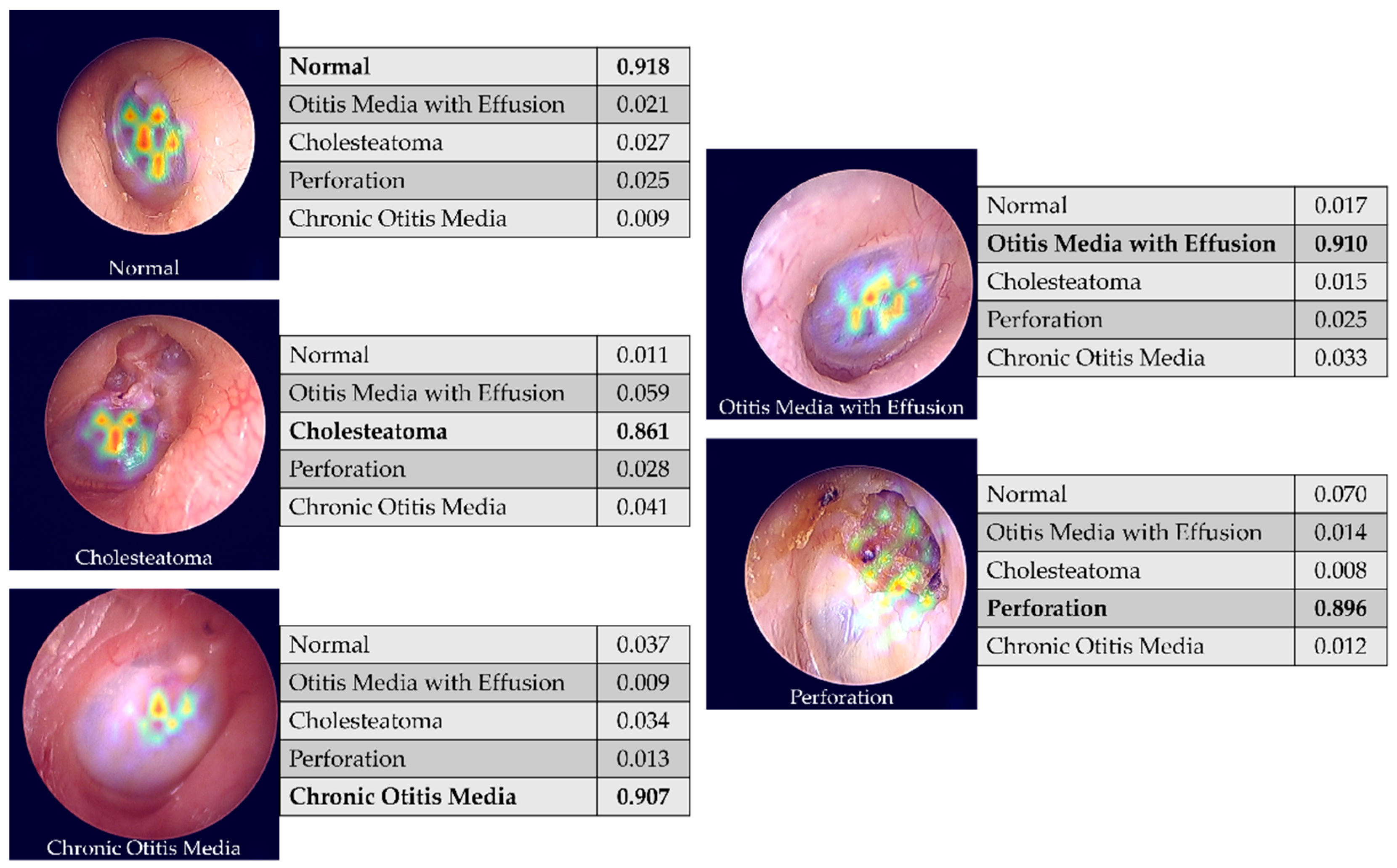
The tympanic membrane itself is a translucent, cone-shaped structure. Because it is semi-transparent, clinicians can see "through" it to the structures of the middle ear, such as the malleus (hammer) and the incus (anvil). When fluid is present, the interface between that fluid and the remaining air creates a visible line or meniscus, much like a spirit level used in construction.
Identifying the Air-Fluid Level: Otoscopic Findings
The hallmark of an air-fluid level is a horizontal line visible behind the tympanic membrane. However, its appearance can vary significantly based on the viscosity of the fluid and the presence of air bubbles.
The Horizontal Meniscus
If the patient is sitting upright, gravity pulls the fluid to the bottom of the middle ear cavity. The surface of this fluid creates a distinct horizontal line. A key diagnostic feature is that this line remains parallel to the ground even if the patient tilts their head forward or backward, provided the fluid is not too viscous.
Air Bubbles (Vesicles)
In many cases, the middle ear is not just "half-full" but contains trapped air bubbles within the fluid. These appear as small, circular outlines behind the drum. Bubbles are highly indicative of a resolving effusion or a state where the Eustachian tube is beginning to function again, intermittently allowing small "gulps" of air to enter the fluid-filled space.
Color and Opacity
The color of the fluid behind the membrane provides clues to the underlying pathology:
- Amber or Straw-Colored: Suggestive of serous otitis media, common in chronic Eustachian tube dysfunction.
- White or Cloudy: Suggestive of mucoid effusion, often seen in "glue ear" (chronic otitis media with effusion).
- Bright Red or Dark Blue/Black: Suggestive of hemotympanum (blood in the middle ear), often following head trauma or severe barotrauma.
- Yellow or Green: Suggestive of purulent fluid associated with acute infection (though in acute infection, the membrane is often too opaque to see a clear fluid level).
Pathophysiology: Why Fluid Accumulates
Fluid behind the eardrum is rarely a primary disease; rather, it is a symptom of an underlying functional or structural issue.
1. Eustachian Tube Dysfunction (ETD)
This is the most common cause. If the tube cannot open, the middle ear tissues absorb the existing oxygen, creating a vacuum. This "ex vacuo" theory suggests that the vacuum then "sucks" fluid out of the capillaries of the mucous membrane. This fluid is typically a transudate (low protein, clear).
2. Inflammation and Infection
During or after an upper respiratory infection, the lining of the middle ear and the Eustachian tube becomes inflamed. This inflammation can block the tube and stimulate goblet cells in the middle ear to produce excess mucus. Even after the infection clears, the mucus may remain, creating an air-fluid level during the recovery phase.
3. Barotrauma
Rapid changes in atmospheric pressure—most commonly during the descent of an aircraft or during scuba diving—can create a sudden, severe pressure differential. If the Eustachian tube cannot equalize this pressure, the resulting vacuum can cause sudden fluid leakage or even the rupture of small blood vessels in the middle ear.
Differential Diagnosis: OME vs. AOM
It is critical to distinguish between Otitis Media with Effusion (OME) and Acute Otitis Media (AOM), as the management of these conditions is vastly different.
| Feature | Otitis Media with Effusion (OME) | Acute Otitis Media (AOM) |
|---|---|---|
| Main Finding | Air-fluid level or bubbles visible. | Bulging, opaque, or erythematous (red) membrane. |
| Pain | Minimal to none; may feel "fullness." | Severe, acute ear pain (otalgia). |
| Fever | Usually absent. | Commonly present. |
| Tympanic Membrane | Neutral or retracted position. | Bulging outward toward the examiner. |
| Treatment | Watchful waiting; addressing ETD. | Antibiotics (if criteria met) and analgesics. |
| Hearing Loss | Conductive hearing loss (mild to moderate). | Conductive hearing loss (often more severe). |
Diagnostic Tools and Procedures
While a standard otoscope is the most common tool, specialized techniques are often required to confirm the presence of fluid when the air-fluid level is not immediately obvious.
Pneumatic Otoscopy
Pneumatic otoscopy is the gold standard for clinical diagnosis of middle ear effusion. By using a rubber bulb to puff air into the ear canal, the clinician observes the movement of the tympanic membrane.
- Normal: The membrane moves briskly in response to pressure.
- Effusion: The presence of fluid behind the membrane significantly dampens or eliminates its movement. If an air-fluid level is present, the fluid itself may be seen to shift or "slosh" slightly under pressure.
Tympanometry
Tympanometry provides an objective measurement of middle ear function. A probe is inserted into the ear canal, and air pressure is varied.
- Type A: Normal middle ear pressure and mobility.
- Type B (Flat): Suggests fluid behind the eardrum (effusion). There is no peak because the fluid prevents the membrane from vibrating.
- Type C: Indicates significant negative pressure in the middle ear, often a precursor to or a resolving stage of effusion.
Audiometry
If an air-fluid level persists for more than a few weeks, an audiogram is necessary to quantify the degree of conductive hearing loss. Fluid acts as a physical barrier to sound conduction, often resulting in a 20 to 40 decibel (dB) hearing loss, which can significantly impact speech development in children.
Clinical Implications and Symptoms
A patient with an air-fluid level behind the tympanic membrane will typically present with a specific cluster of symptoms. Understanding these helps the clinician correlate the visual findings with the patient's lived experience.
- Aural Fullness: Patients often describe a sensation of "water in the ear" or a "clogged" feeling that doesn't resolve with yawning.
- Conductive Hearing Loss: Sound may seem muffled, as if the patient is "underwater."
- Autophony: The patient's own voice may sound unusually loud or distorted in the affected ear.
- Popping or Crackling: As the Eustachian tube attempts to open, the air moving into the fluid creates popping sounds, which are often a sign that the condition is beginning to resolve.
- Balance Issues: While less common than hearing issues, the presence of fluid can occasionally cause mild vertigo or a sense of unsteadiness, as the middle ear plays a role in the overall sensory input for balance.
Management and Treatment Strategies
As of 2026, clinical guidelines emphasize a conservative approach to air-fluid levels associated with OME, while being more aggressive with barotrauma or chronic cases.
1. Watchful Waiting
Most cases of OME resolve spontaneously within three months. If the patient is not at high risk (e.g., no significant developmental delays or severe bilateral hearing loss), clinicians typically monitor the fluid every 4 to 8 weeks.
2. Treating the Underlying Cause
- Allergy Management: If the Eustachian tube is blocked due to allergic rhinitis, intranasal corticosteroids or antihistamines may help reduce inflammation at the tube's opening.
- Decongestants: While not recommended for long-term use, short-term oral or nasal decongestants may be used in adults to help open the Eustachian tube during a viral infection.
- Valsalva Maneuver: Patients may be instructed to gently "pop" their ears to force air into the middle ear, helping to clear the fluid.
3. Surgical Intervention
If the air-fluid level persists for more than three months (chronic OME) or if it causes significant hearing impairment, surgical options are considered:
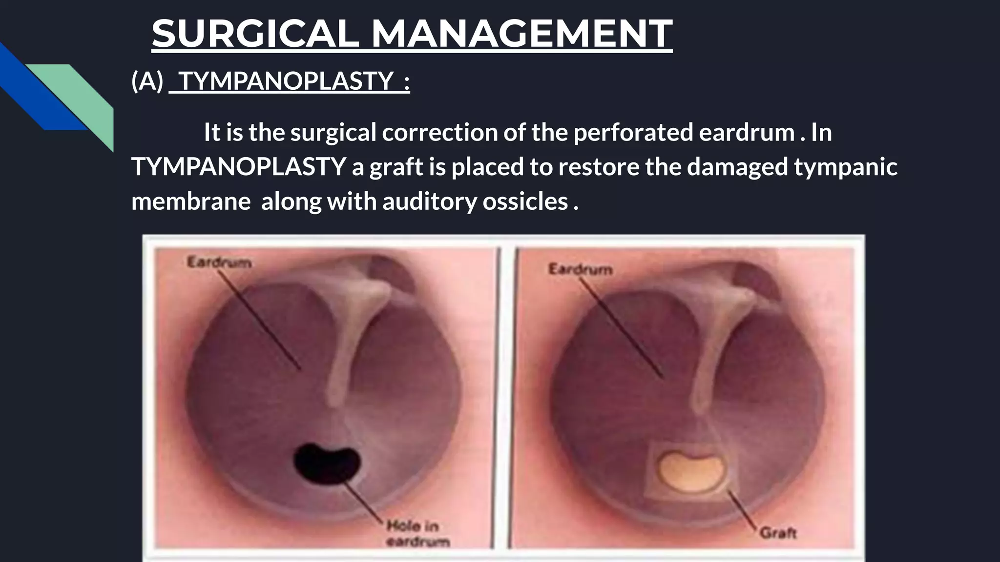
- Myringotomy: A small incision is made in the tympanic membrane to drain the fluid.
- Tympanostomy Tubes (PE Tubes): Small tubes are placed in the eardrum to provide a constant source of ventilation, bypassing the dysfunctional Eustachian tube. This is the most common surgical procedure in children in the United States.
Complications of Untreated Effusion
While an air-fluid level often resolves on its own, chronic fluid can lead to more serious conditions:
- Tympanic Membrane Retraction: Chronic negative pressure can pull the eardrum inward, potentially damaging the ossicular chain.
- Cholesteatoma: A skin cyst that can form in a retraction pocket, which can eventually erode the bones of the middle ear.
- Tympanosclerosis: Scarring of the eardrum, which can lead to permanent stiffness and mild hearing loss.
- Speech and Language Delay: In children, the persistent muffled hearing caused by fluid can lead to delays in reaching developmental milestones.
Frequently Asked Questions
Can I fly with an air-fluid level in my ear?
Flying with an active middle ear effusion or an air-fluid level is generally safer than flying with a completely blocked Eustachian tube that has no fluid yet. However, the pressure changes during descent can be painful. It is recommended to use a nasal decongestant 30 minutes before descent and practice the Valsalva maneuver. If the fluid is a result of recent barotrauma, consult a physician before flying again.
How long does it take for fluid behind the eardrum to disappear?
In most cases following a cold or an episode of acute otitis media, fluid will resolve within 2 to 4 weeks. However, approximately 10% of children will still have fluid after 3 months. If an air-fluid level is still visible after 12 weeks, a referral to an Ear, Nose, and Throat (ENT) specialist is usually warranted.
Does an air-fluid level mean I have an ear infection?
Not necessarily. An air-fluid level is the primary sign of Otitis Media with Effusion (OME), which is often not an active bacterial infection. OME is the accumulation of sterile fluid. While it can follow an infection, the presence of a fluid level without pain or fever usually means antibiotics are not required.
Can allergies cause an air-fluid level?
Yes, allergies are a leading cause of Eustachian tube dysfunction. Chronic inflammation of the nasal passages and nasopharynx can block the Eustachian tube, leading to the negative pressure that creates an effusion and a visible air-fluid level.
Why do I see bubbles behind my eardrum?
Bubbles are actually a good sign. They indicate that air is successfully making its way through the Eustachian tube and into the fluid-filled middle ear. This usually means the Eustachian tube is starting to function again and the effusion is in the process of resolving.
Practical Recommendation for Patients and Clinicians
If an air-fluid level is detected during an examination, the immediate next step is to assess the patient's hearing and the duration of the symptoms. For adults, a sudden onset of unilateral fluid without a preceding cold should be investigated by an ENT to rule out nasopharyngeal masses blocking the Eustachian tube. For children, the focus should be on monitoring developmental milestones and hearing. In both cases, maintaining nasal hygiene and managing allergies are effective first-line steps to encourage the natural clearance of the fluid.