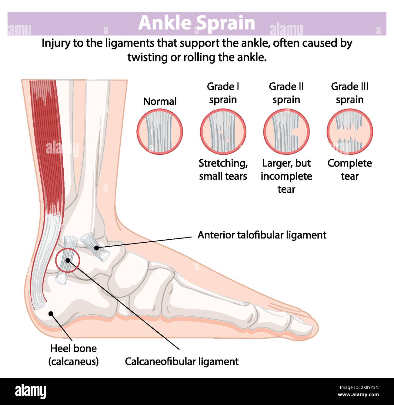
Ankle ligaments are strong, fibrous bands of connective tissue that anchor the bones of the ankle joint together, providing essential stability. They are categorized into three primary groups: the lateral complex, the medial (deltoid) complex, and the syndesmotic ligaments, which facilitate controlled movement while preventing excessive joint displacement.
The human ankle is a complex hinge joint (the talocrural joint) that bears the entire weight of the body while allowing for multi-directional movement. Because the ankle must balance mobility with rigid support, its ligamentous structure is incredibly dense and specialized. Understanding the "ankle ligaments labeled" in a clinical or anatomical context requires looking at the joint from three distinct perspectives: the outer (lateral) side, the inner (medial) side, and the superior (syndesmotic) connection between the lower leg bones.
As of 2026, ligamentous health remains a primary focus of sports medicine and orthopedic surgery. With the rising popularity of high-impact sports and a global push toward active aging, the incidence of ankle sprains and chronic instability has led to more precise diagnostic labeling and advanced rehabilitation protocols.
The Lateral Ligament Complex (Outer Ankle)
The lateral side of the ankle is the most frequent site of injury, specifically during "inversion" events where the foot rolls inward. The lateral ligament complex consists of three distinct bands that connect the fibula (the smaller outer leg bone) to the talus and calcaneus (heel bone).
Anterior Talofibular Ligament (ATFL)
The ATFL is the most commonly injured ligament in the entire human body. It is a short, flat band that extends from the anterior margin of the lateral malleolus (the bony bump on the outer ankle) to the neck of the talus. Biomechanically, the ATFL is responsible for resisting internal rotation of the talus and preventing the foot from sliding forward relative to the leg.
In clinical labeling, the ATFL is often the first point of assessment. Because it is at its tightest when the foot is pointed downward (plantarflexion), it is highly susceptible to tearing when an individual "turns" their ankle while walking or running.
Calcaneofibular Ligament (CFL)
Located just below the ATFL, the CFL is a cord-like band that connects the tip of the lateral malleolus to a small tubercle on the outer surface of the calcaneus. Unlike the ATFL, the CFL crosses two joints: the talocrural joint and the subtalar joint. This dual-joint coverage makes it vital for stabilizing the heel during side-to-side movements.
In cases of severe inversion sprains, the CFL is typically the second ligament to tear after the ATFL. When both are compromised, the joint loses significant lateral stability, often leading to a "wobbly" sensation during gait.
Posterior Talofibular Ligament (PTFL)
The PTFL is the strongest and deepest of the three lateral ligaments. it runs almost horizontally from the posterior (back) part of the lateral malleolus to the posterior process of the talus. Due to its thickness and position, it is rarely injured in isolation. It generally only sustains damage during total ankle dislocations or extremely high-force trauma. Its primary role is to prevent the talus from shifting backward and to provide stability when the foot is flexed upward (dorsiflexion).
The Medial Ligament Complex (The Deltoid Ligament)
On the inner side of the ankle lies the deltoid ligament, a massive, triangular-shaped structure that is significantly stronger than the lateral ligaments. Because the deltoid ligament is so robust and the medial malleolus (inner ankle bone) is physically shorter than the lateral side, medial ankle sprains (eversion sprains) are much less common, accounting for less than 10% of all ankle injuries.
The deltoid ligament is divided into two layers: the superficial and the deep components.
Superficial Layer
The superficial deltoid ligament consists of three main parts that originate from the medial malleolus and fan out to different bones in the midfoot and heel:
- Tibionavicular Ligament: Connects the tibia to the navicular bone.
- Tibiocalcaneal Ligament: Connects the tibia to the sustentaculum tali of the calcaneus.
- Superficial Posterior Tibiotalar Ligament: Connects the tibia to the back of the talus.
These bands work together to prevent the foot from rolling outward (eversion) and to stabilize the longitudinal arch of the foot.
Deep Layer
The deep layer is composed of the Anterior Tibiotalar Ligament and the Deep Posterior Tibiotalar Ligament. These are arguably the most important stabilizers of the medial ankle, preventing the talus from shifting laterally or rotating excessively within the ankle mortise. Because these ligaments are so deep and strong, a force great enough to tear them often results in a fracture of the medial malleolus bone instead of a pure ligamentous rupture.
The Syndesmotic Ligament Complex (The High Ankle)
The "high ankle" refers to the syndesmosis, the fibrous joint that holds the tibia (shin bone) and fibula together just above the ankle joint. This area is critical for maintaining the "mortise"—the socket-like structure where the talus sits. If the syndesmosis is compromised, the two leg bones can spread apart, making the ankle joint unstable and painful.
Components of the Syndesmosis
- Anterior Inferior Tibiofibular Ligament (AITFL): Located on the front of the ankle, this is the ligament most commonly injured in a "high ankle sprain." It resists the external rotation of the foot.
- Posterior Inferior Tibiofibular Ligament (PITFL): Located on the back, this ligament is very thick and strong, providing posterior stability to the lower leg bones.
- Transverse Ligament: A lower extension of the PITFL that helps create a shelf for the talus.
- Interosseous Membrane and Ligament: A vast sheet of connective tissue that runs between the entire length of the tibia and fibula, acting as a massive stabilizer.
High ankle sprains are particularly difficult to treat because every time a person stands up, their body weight naturally tries to push the tibia and fibula apart. Consequently, the healing time for syndesmotic injuries is often twice as long as that for standard lateral sprains.
Summary of Ankle Ligaments and Functions
The following table provides a quick reference for the primary ligaments discussed, their locations, and their clinical significance in 2026.
| Ligament Group | Specific Ligament | Location | Primary Role | Common Injury Name |
|---|---|---|---|---|
| Lateral | ATFL | Outer Front | Resists inversion / internal rotation | Standard Ankle Sprain |
| Lateral | CFL | Outer Side | Stabilizes heel and subtalar joint | Grade II/III Sprain |
| Lateral | PTFL | Outer Back | Resists posterior talar displacement | Severe Trauma / Dislocation |
| Medial | Deltoid (Superficial) | Inner Side | Resists eversion (outward roll) | Eversion Sprain |
| Medial | Deltoid (Deep) | Inner Deep | Primary medial talar stabilizer | Medial Malleolus Fracture |
| Syndesmosis | AITFL | Lower Leg Front | Holds tibia and fibula together | High Ankle Sprain |
| Syndesmosis | Interosseous | Between Bones | Prevents widening of the mortise | Severe High Ankle Injury |
Identifying and Grading Ligament Injuries
When ankle ligaments are labeled in a medical report, they are usually accompanied by a "grade" that describes the severity of the damage. As of 2026, the standard three-tier grading system remains the most effective tool for guiding treatment.
Grade I: Mild Sprain
A Grade I injury involves microscopic tearing or stretching of the ligament fibers, most commonly the ATFL. There is usually minimal swelling and no joint instability. Patients can typically bear weight, though they may experience soreness. Recovery usually takes 1 to 3 weeks.
Grade II: Moderate Sprain
This involves a partial tear of the ligament. In lateral injuries, this often means a complete tear of the ATFL and a partial tear of the CFL. There is significant swelling, bruising (ecchymosis), and moderate pain. The joint may feel slightly loose during clinical testing (such as the "Anterior Drawer Test"). Recovery often requires 4 to 8 weeks and physical therapy.
Grade III: Severe Sprain
A Grade III injury is a complete rupture of one or more ligaments. If the lateral side is involved, both the ATFL and CFL are typically fully torn, and the PTFL may be damaged. The ankle is unstable, weight-bearing is often impossible, and the swelling is intense. While many Grade III injuries can be managed with rigorous rehabilitation, some may require surgical intervention to restore stability, especially in athletes.
Diagnostic Procedures for Labeled Ligament Analysis
In 2026, healthcare providers use a combination of physical exams and advanced imaging to identify which ankle ligaments are damaged.
- Physical Examination: Special tests like the Anterior Drawer Test (for ATFL) and the Talar Tilt Test (for CFL) allow doctors to feel for "laxity" or excessive movement in the joint.
- X-rays: While X-rays do not show ligaments, they are essential for ruling out "avulsion fractures," where a ligament pulls a small piece of bone away during an injury. Stress X-rays may also be used to see if the joint gapes under pressure.
- MRI (Magnetic Resonance Imaging): This is the gold standard for visualizing soft tissue. An MRI provides clear, labeled images of every ligament, allowing radiologists to see the exact location and grade of a tear.
- Musculoskeletal Ultrasound: This has become increasingly popular in 2026 for point-of-care diagnostics. It is a cost-effective, dynamic way to see how the ligaments behave while the ankle is in motion.
Rehabilitation and Recovery Protocols
The goal of treating injured ankle ligaments is to prevent "Chronic Ankle Instability" (CAI). When a ligament heals "long" or becomes lax, the mechanoreceptors within the tissue—which tell the brain where the foot is in space—become impaired.
The R.I.C.E. and P.O.L.I.C.E. Methods
While the traditional R.I.C.E. (Rest, Ice, Compression, Elevation) method is still used, modern 2026 guidelines favor the P.O.L.I.C.E. protocol:
- Protection (using a brace or boot)
- Optimal Loading (gradual weight-bearing to encourage tissue healing)
- Ice
- Compression
- Elevation
Physical Therapy Focus
Rehabilitation focuses on three main pillars:
- Range of Motion: Restoring the ability to move the foot up and down without pain.
- Strengthening: Specifically targeting the peroneal muscles on the side of the leg, which act as "dynamic stabilizers" to support the lateral ligaments.
- Proprioception: Exercises using balance boards or foam pads to retrain the nervous system to stabilize the joint during unpredictable movements.
Surgical Options for Chronic Instability
For patients whose ligaments do not heal properly, the Broström-Gould procedure is the most common surgical intervention. In this surgery, the surgeon tightens the lateral ligaments (ATFL and CFL) and may reinforce them using the extensor retinaculum (a nearby band of tissue). In 2026, many of these procedures are performed arthroscopically, leading to smaller incisions and faster recovery times.
For more information on the anatomy of the foot and ankle, you can consult the American Academy of Orthopaedic Surgeons (AAOS) or view detailed anatomical breakdowns on StatPearls via the NCBI.
Frequently Asked Questions
Which ankle ligament is most commonly torn?
The Anterior Talofibular Ligament (ATFL) is the most frequently injured ligament in the ankle. It is located on the outer front of the joint and is the primary restraint against inversion. Because it is the weakest of the lateral complex and is under the most tension when the foot is pointed, it is typically the first structure to fail during a common ankle sprain.
What is a "high ankle" ligament?
The "high ankle" ligaments are collectively known as the syndesmosis. They include the AITFL, PITFL, and the interosseous membrane. These ligaments sit above the actual hinge of the ankle and hold the tibia and fibula together. Injuries here are usually caused by the foot being forced upward and outward (external rotation), and they generally require a significantly longer recovery period than standard sprains.
Can a torn ankle ligament heal without surgery?
Yes, the vast majority of ankle ligament tears, including Grade III ruptures, can heal without surgery. The body is capable of repairing these fibrous tissues through the formation of scar tissue. However, successful non-surgical healing depends heavily on proper immobilization followed by a structured physical therapy program to restore strength and balance. Surgery is typically reserved for elite athletes or those with chronic instability that does not respond to therapy.
How do I know if I injured the deltoid ligament?
A deltoid ligament injury occurs on the inner (medial) side of the ankle. You will likely experience pain, swelling, and bruising on the inner bony prominence. These injuries are often the result of an eversion force—where the bottom of the foot turns outward. Because these injuries are often associated with fractures, a clinical evaluation and X-rays are highly recommended if you experience significant medial ankle pain.
What is the difference between a ligament and a tendon in the ankle?
A ligament, such as the ATFL, connects bone to bone and provides passive stability to the joint. A tendon, such as the Achilles or the Peroneal tendons, connects muscle to bone and allows for active movement by transmitting force from muscle contractions. While both are made of collagen, they serve different functional roles and require different rehabilitation approaches when injured.
Next Actions for Ankle Health
If you suspect you have injured an ankle ligament, the first step is to follow the P.O.L.I.C.E. protocol to manage inflammation. Seek a professional evaluation from a physical therapist or orthopedic specialist to determine the specific ligaments involved. Early diagnosis and "optimal loading" are the best ways to ensure a full recovery and prevent the long-term complication of chronic ankle instability. For those looking to prevent future injuries, incorporating daily balance exercises (single-leg stands) can significantly strengthen the neural pathways that protect your ankle ligaments.