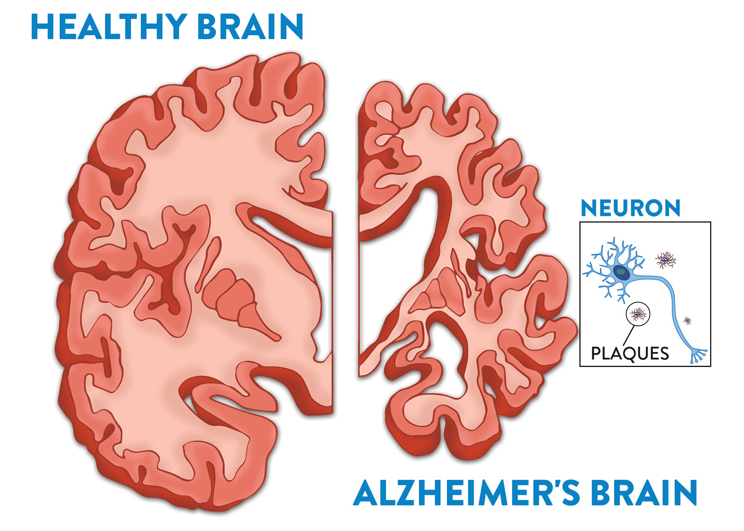
The primary difference between an Alzheimer’s brain and a regular brain lies in the physical structure, cellular health, and chemical signaling. While a healthy brain maintains its weight and volume with efficient neuronal communication, an Alzheimer’s brain suffers from severe atrophy, the accumulation of toxic protein aggregates (plaques and tangles), and widespread cell death that disrupts memory and cognition.
Understanding the distinction between normal age-related changes and the pathological decline of Alzheimer’s disease is essential for early intervention and care planning. As of 2026, neuroscientists have mapped the progression of the disease with unprecedented detail, highlighting that the "Alzheimer's brain" is not merely an accelerated version of an aging brain, but a fundamentally different biological state.
The Biological Blueprint: How a Healthy Brain Functions
To understand the devastation of Alzheimer’s, one must first appreciate the complexity of a regular, healthy brain. The human brain contains approximately 86 billion neurons—specialized cells that serve as the fundamental units of the nervous system. These neurons communicate through electrical impulses and chemical signals called neurotransmitters.
In a healthy brain, synapses (the gaps between neurons) are clear and functional. Information travels across these gaps seamlessly, allowing for the formation of memories, the execution of movements, and the regulation of emotions. A regular brain also possesses a highly efficient waste-clearance system, often referred to as the glymphatic system, which flushes out metabolic byproducts—including potentially harmful proteins—during sleep.
While a regular brain does experience some shrinkage as a person reaches their 70s and 80s, this "normal aging" is typically subtle. The brain loses some volume, and processing speed may slow slightly, but the fundamental architecture remains intact. Critical regions like the hippocampus, which handles new memory formation, stay relatively robust in healthy aging, allowing individuals to maintain their independence and cognitive core.
The Microscopic Shift: Amyloid Plaques and Tau Tangles
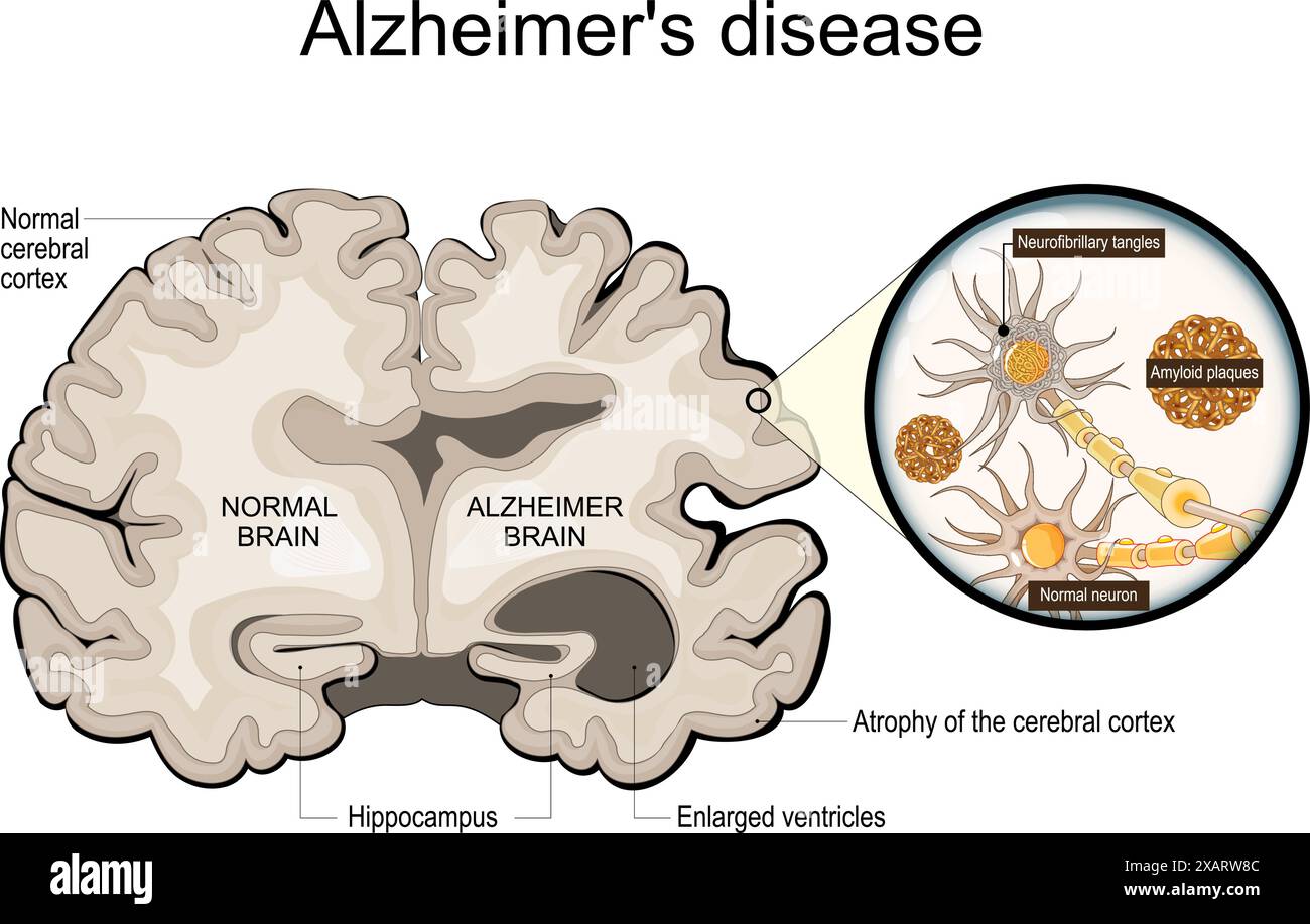
The most significant microscopic difference in an Alzheimer’s brain vs. a regular brain is the presence of two specific protein abnormalities: extracellular amyloid plaques and intracellular neurofibrillary tangles.
Beta-Amyloid Plaques
In a healthy brain, beta-amyloid protein fragments are produced and normally broken down or eliminated. In an Alzheimer’s brain, these fragments clump together to form hard, insoluble plaques between neurons. These plaques interfere with the synapses, effectively "clogging" the communication lines. By 2026, research has confirmed that amyloid accumulation often begins 15 to 20 years before the first symptoms of memory loss appear. These plaques are toxic to neurons and trigger an immune response that, over time, causes more harm than good.
Neurofibrillary Tangles (Tau)
While plaques happen outside the cells, tangles happen inside. In a regular brain, a protein called tau helps stabilize microtubules—the internal structures that transport nutrients and molecules within the neuron. In Alzheimer’s disease, the tau protein undergoes chemical changes that cause it to detach from microtubules and stick to other tau molecules, forming threads that eventually tie into tangles. When these tangles form, the neuron's transport system collapses. Imagine a train track falling apart; the "supplies" can no longer reach the cell, and the neuron eventually dies.
Structural Atrophy: Measuring the Physical Decline
When comparing an Alzheimer’s brain to a regular brain through imaging such as MRI, the physical differences are stark. Alzheimer’s disease causes the brain to shrink significantly, a process known as atrophy.
- The Hippocampus: This is often the first area to show significant damage. In a regular brain, the hippocampus is plump and functional. In the Alzheimer’s brain, it shrivels, which explains why short-term memory loss is usually the first clinical symptom.
- The Cerebral Cortex: This outer layer is responsible for high-level functions like language, reasoning, and social behavior. As neurons die, the cortex thins out. This leads to the characteristic "gaps" or widened furrows (sulci) seen in brain scans of patients.
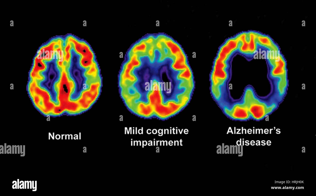
- The Ventricles: These are fluid-filled spaces inside the brain. In a regular brain, they are small and contained. As the surrounding brain tissue dies in Alzheimer’s, the ventricles expand to fill the void, appearing much larger on an MRI.
According to the National Institute on Aging, by the final stages of the disease, the brain's weight can decrease by as much as one-third compared to a healthy brain of the same age.
Chemical Communication: Neurotransmitter Disruptions
A regular brain maintains a delicate balance of chemicals to ensure smooth operation. In Alzheimer’s, this balance is destroyed. The most notable deficit occurs with acetylcholine, a neurotransmitter essential for learning and memory.
Neurons that produce acetylcholine are among the first to be destroyed by the disease. This is why many of the standard treatments available in 2026, such as cholinesterase inhibitors, focus on preventing the breakdown of the remaining acetylcholine to temporarily boost communication between surviving neurons.
Additionally, the Alzheimer’s brain often suffers from an overabundance of glutamate. While glutamate is necessary for signaling, too much of it can overstimulate neurons, leading to "excitotoxicity"—essentially, the cells are worked to death. This chemical chaos is absent in a regular aging brain, where neurotransmitter levels remain within a functional range.
Metabolic Slowdown: Glucose Utilization and Energy Failure
The brain is an energy-intensive organ, consuming about 20% of the body’s total oxygen and glucose. A regular brain is highly efficient at converting glucose into energy to power neuronal firing.
In contrast, the Alzheimer’s brain exhibits a condition often described as "type 3 diabetes" or brain insulin resistance. Using PET scans (Positron Emission Tomography), doctors can observe that the Alzheimer's brain has a significantly reduced ability to uptake glucose. This metabolic failure means that even if the neurons are still alive, they lack the "fuel" necessary to function correctly. This energy crisis often precedes the death of the cells and contributes to the cognitive "fog" and confusion experienced by patients.
Inflammation and the Immune Response
In a regular brain, immune cells called microglia act as the primary defenders, clearing away debris and pathogens. They are the "janitors" of the central nervous system. In a healthy state, they perform their duties and then return to a resting state.
In an Alzheimer’s brain, the microglia become chronically activated. Because they cannot efficiently clear the massive buildup of amyloid plaques, they remain in a state of high alert, releasing pro-inflammatory cytokines. Instead of protecting the brain, this chronic inflammation begins to damage healthy neurons. This shift from "protector" to "aggressor" is a hallmark of the Alzheimer's pathology and is a major focus of therapeutic research in 2026.
Comparison: Healthy Aging vs. Alzheimer’s Disease
The following table summarizes the primary distinctions between the structural and functional aspects of a regular aging brain and one affected by Alzheimer's.
| Feature | Regular Aging Brain | Alzheimer's Disease Brain |
|---|---|---|
| Brain Volume | Slight, gradual shrinkage over decades. | Rapid, severe atrophy and weight loss. |
| Protein Deposits | Minimal to no amyloid or tau buildup. | Extensive plaques and neurofibrillary tangles. |
| Hippocampus | Largely preserved; mild memory slowing. | Severe shriveling; inability to form new memories. |
| Ventricles | Maintain standard size for age. | Significant enlargement (ventriculomegaly). |
| Cell Communication | Synapses remain mostly intact. | Synaptic loss; breakdown in neurotransmission. |
| Glucose Metabolism | Efficient energy conversion. | Impaired; cells struggle to use glucose for fuel. |
| Waste Clearance | Functional glymphatic system. | Failed clearance leading to toxic accumulation. |
Functional Consequences: Memory, Language, and Executive Function
The physical differences translate directly into the symptoms observed in patients. In a regular brain, a person might occasionally forget a name or misplace their keys but can usually retrace their steps or remember the information later. This is a retrieval issue, not a storage issue.
In an Alzheimer’s brain, the information is often never "stored" in the first place because the hippocampus is too damaged to encode it. As the disease spreads from the hippocampus to the temporal and parietal lobes, language becomes affected. A person with a regular brain might experience "tip-of-the-tongue" moments; a person with Alzheimer's might lose the ability to speak or understand complex sentences entirely.
Eventually, the damage reaches the frontal lobe, which governs executive function—the ability to plan, solve problems, and control impulses. While a healthy older adult can manage their finances and schedule, an Alzheimer's patient loses the "map" for these complex tasks as the frontal neurons disconnect and die.
Diagnostic Tools in 2026: Visualizing the Difference
As of 2026, the ability to distinguish an Alzheimer's brain from a regular brain has moved beyond post-mortem examination. Clinical diagnostics now utilize a multi-modal approach to catch the disease early:
- Blood-Based Biomarkers: High-sensitivity tests for p-tau217 and amyloid-beta 42/40 ratios allow doctors to detect brain changes from a simple blood draw, often with 90% accuracy.
- Advanced PET Imaging: New tracers can now visualize tau tangles in real-time, helping clinicians see exactly where the disease is spreading.
- Volumetric MRI: Software can now automatically calculate the volume of the hippocampus and compare it to a normative database of "regular" brains to identify abnormal shrinkage.
These tools are vital because, while many symptoms of Alzheimer's overlap with other forms of dementia or even severe depression, the biological signature of the Alzheimer's brain—the plaques and tangles—is unique.
Common Questions Regarding Brain Health
Can you prevent a regular brain from becoming an Alzheimer's brain?
While genetics (such as the APOE-ε4 allele) play a role, lifestyle factors significantly influence risk. Regular cardiovascular exercise, a Mediterranean-style diet, and managing blood pressure are proven to support the glymphatic system and reduce neuroinflammation. However, there is currently no guaranteed "prevention" for the underlying pathology.
Is brain shrinkage always a sign of Alzheimer's?
No. Some brain shrinkage is a normal part of aging. Furthermore, other conditions like chronic alcoholism, certain vitamin deficiencies, and chronic stress can cause atrophy. The key difference in Alzheimer’s is the pattern and speed of the shrinkage, combined with the presence of specific protein markers.
Does a regular brain have any plaques at all?
Some cognitively healthy older adults do show small amounts of amyloid plaques in their brains. This is often referred to as "preclinical Alzheimer's." Research suggests these individuals may be at high risk for future decline, but their brains are currently compensating for the damage, whereas an Alzheimer's-diagnosed brain has reached a threshold where compensation is no longer possible.
How does sleep affect the difference between the two?
Sleep is the "cleaning cycle" for the brain. During deep sleep, the space between brain cells increases, allowing cerebrospinal fluid to wash away beta-amyloid. A regular brain typically benefits from consistent, quality sleep. Chronic sleep deprivation is linked to higher amyloid levels, potentially pushing a regular brain toward an Alzheimer's state.
Can the damage in an Alzheimer's brain be reversed?
As of 2026, most treatments focus on slowing the progression or clearing amyloid plaques (using monoclonal antibodies). While these can slow the decline, they generally do not "regrow" dead neurons or restore the brain to its "regular" pre-disease state. Early detection remains the most effective way to preserve existing function.
Recommendations for Monitoring Brain Health
If you are concerned about the differences between your cognitive function and what is considered "normal," the first step is a baseline cognitive assessment. In 2026, these are often performed digitally and can track minor fluctuations in memory and executive function over time.
For those with a family history of neurodegenerative disease, participating in longitudinal studies or seeking a referral to a neurologist for biomarker testing can provide clarity. Understanding that the Alzheimer's brain is a distinct biological entity—not an inevitable part of growing older—is the first step toward proactive brain health management. Maintaining cardiovascular health, staying socially active, and engaging in lifelong learning remain the most effective ways to support a regular, healthy brain well into your later years.