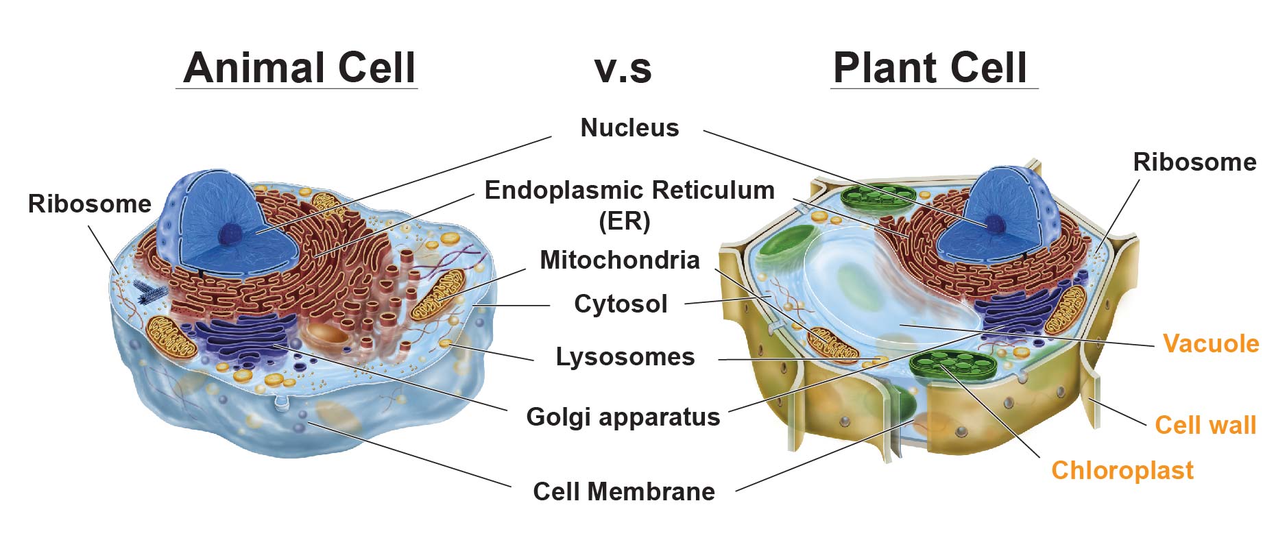
Plant and animal cells differ primarily in that plant cells contain a rigid cell wall surrounding their plasma membrane, large central vacuoles for turgor pressure regulation, and chloroplasts capable of photosynthesis, whereas animal cells lack these structures and rely instead on flexible extracellular matrices and smaller, transient vacuoles.
Understanding the fundamental differences between plant and animal cells is a cornerstone of cellular biology. While both are eukaryotic cells containing membrane-bound nuclei and sophisticated organelles, their distinct structural components reflect the unique physiological demands and evolutionary paths of the kingdoms they belong to. Plants require rigid support against gravity and self-sustaining energy production via sunlight, while animals require mobility, structural flexibility, and consumption-based metabolism. Examining these microscopic units reveals how structural variations translate directly into macroscopic life functions.
Foundational Overview of Eukaryotic Cell Architecture
To understand the distinctions between plant and animal cells, one must first recognize their shared lineage. Both plant cells (from the kingdom Plantae) and animal cells (from the kingdom Animalia) are classified as eukaryotic cells. This means they share a complex internal architecture characterized by a true nucleus housing linear DNA, a cytoskeleton providing structural organization, and a suite of endomembrane-bound organelles such as the endoplasmic reticulum, Golgi apparatus, mitochondria, and ribosomes.
Despite this shared foundation, evolutionary pressures forced divergent adaptations. Animals evolved mobility, heterotrophic nutrition (obtaining energy by consuming other organisms), and specialized nervous and muscular systems. Plants evolved autotrophic nutrition (producing their own food via photosynthesis), sessile lifestyles (anchored to a single location), and exposure to variable environmental conditions like drought, intense solar radiation, and wind shear. These evolutionary trajectories shaped the three primary structural and functional divisions that distinguish plant cells from animal cells.
Distinction 1: The Cell Wall Versus the Flexible Plasma Membrane
The most immediate visual and mechanical difference under a light microscope is the boundary layer of the cell. While both plant and animal cells possess a phospholipid bilayer known as the plasma (or cell) membrane, plant cells feature an additional, highly rigid extracellular matrix called the cell wall located exterior to the plasma membrane.
Composition and Architecture of the Plant Cell Wall
The plant cell wall is composed primarily of cellulose microfibrils embedded in a complex matrix of hemicellulose and pectins. When a plant cell first divides, it secretes a thin, sticky layer called the middle lamella that glues adjacent cells together. It then builds a primary cell wall, which is flexible enough to allow for cell growth. In many mature plant cells, a secondary cell wall is deposited inside the primary wall, containing lignin—a rigid, waterproof polymer that provides immense structural strength.
Functional Implications for Plants
- Structural Support: The rigidity of the cell wall allows plants to grow upright against the force of gravity without requiring a skeletal system.
- Turgor Pressure Regulation: The wall acts as a containment vessel, preventing the cell from bursting when water enters via osmosis. This internal pressure, known as turgor pressure, keeps non-woody plant stems crisp and leaves turgid.
- Pathogen Defense: The thick matrix serves as a physical barrier against bacteria, fungi, and mechanical damage.
The Animal Alternative: Extracellular Matrix
Animal cells completely lack a cell wall. Their outermost layer is the flexible plasma membrane, which is supplemented on the outside by a diverse network of glycoproteins, collagen, and proteoglycans known as the extracellular matrix (ECM). While the ECM provides structural support, tissue adhesion, and cell signaling capabilities, it lacks the rigid tensile strength of cellulose, allowing animal tissues to remain flexible, contractile, and capable of complex movement.
| Feature | Plant Cell Boundary | Animal Cell Boundary |
|---|---|---|
| Outermost Layer | Rigid cell wall (cellulose, hemicellulose, pectin) | Flexible plasma membrane with Extracellular Matrix (ECM) |
| Rigidity | High; provides fixed shape and support | Low to moderate; allows flexibility and motility |
| Osmotic Protection | High resistance to osmotic lysis via cell wall | Susceptible to lysis if internal water volume is unregulated |
| Intercellular Connection | Plasmodesmata (channels through walls) | Gap junctions, tight junctions, and desmosomes |
Distinction 2: Plastids and Chloroplasts for Energy Production
Energy acquisition is another major dividing line between plant and animal cellular physiology. While both cell types house mitochondria—the powerhouses responsible for cellular respiration and ATP generation—plant cells possess an additional class of organelles called plastids, most notably chloroplasts.
Structure and Function of Chloroplasts
Chloroplasts are specialized, double-membrane-bound organelles containing an internal system of disc-like membranes called thylakoids, stacked into structures known as grana, suspended in a fluid matrix called the stroma. Chloroplasts contain the green pigment chlorophyll, which captures light energy from the sun.
Through the process of photosynthesis, plant cells convert carbon dioxide and water into glucose and oxygen, utilizing solar energy. This autotrophic capability means plant cells manufacture their own nutritional fuel internally. Other types of plastids, such as leucoplasts and chromoplasts, store starches, lipids, or synthesize pigments for flowers and fruits.
The Animal Heterotrophic Approach
Animal cells possess no chloroplasts or plastids. Because animals are heterotrophs, their cells cannot fix carbon dioxide into organic molecules using light energy. Instead, animal cells rely exclusively on consuming organic matter. Mitochondria inside animal cells break down these consumed carbohydrates, fats, and proteins through cellular respiration to harvest chemical energy in the form of adenosine triphosphate (ATP).
While plant cells also rely heavily on mitochondria at night or in non-green tissues (like roots), their ability to generate their own sugars via chloroplasts during daylight hours represents a fundamental metabolic divergence from animal cells.
Distinction 3: Vacuolar Systems (Large Central Vacuole vs. Small Vacuoles)
The management of internal storage, waste disposal, and turgor pressure relies on vacuoles—membrane-bound fluid-filled sacs. Here, the structural difference between plant and animal cells is starkly apparent in both size and function.
The Plant Large Central Vacuole
Mature plant cells typically contain a single, massive central vacuole that can occupy up to 90% of the cell's total volume. Pushed against the cell wall by this giant organelle, the cytoplasm forms a thin peripheral layer.
The central vacuole is filled with cell sap—a watery solution containing ions, sugars, enzymes, amino acids, and waste products. Its primary functions include:
- Turgor Pressure Maintenance: By absorbing or releasing water, the central vacuole regulates internal pressure against the cell wall, driving cell elongation and preventing wilting.
- Storage and Isolation: It stores vital nutrients and sequesters toxic metabolic byproducts away from the rest of the cellular machinery.
- Enzymatic Degradation: It contains hydrolytic enzymes that perform functions similar to animal lysosomes.
Animal Vacuolar Systems
Animal cells do not possess a large central vacuole. Instead, they contain multiple, much smaller, temporary vacuoles scattered throughout the cytoplasm. These small vacuoles are primarily involved in endocytosis (taking substances into the cell) and exocytosis (expelling waste). Furthermore, animal cells rely on specialized organelles called lysosomes—which contain digestive enzymes to break down waste and cellular debris—a role largely handled by the central vacuole and specialized vesicles in plants.
Structural Comparison Summary
To synthesize these differences clearly, the following matrix contrasts the key structural, metabolic, and architectural attributes of plant and animal cells.
| Cellular Component | Plant Cells | Animal Cells |
|---|---|---|
| Cell Wall | Present (rigid, made of cellulose) | Absent |
| Chloroplasts / Plastids | Present (site of photosynthesis) | Absent |
| Central Vacuole | One large central vacuole (up to 90% volume) | Multiple small, temporary vacuoles |
| Shape | Fixed, regular rectangular or polygonal shape | Irregular, variable, and flexible shape |
| Lysosomes | Rare (functions often handled by vacuoles) | Generally present and abundant |
| Centrioles / Centrosomes | Absent in most higher plants (present in lower plants) | Present in most cell types |
| Energy Storage | Starch | Glycogen |
| Intercellular Channels | Plasmodesmata | Gap junctions |
Frequently Asked Questions
Do plant cells have mitochondria?
Yes. A common misconception is that because plant cells have chloroplasts for photosynthesis, they do not need mitochondria. Plant cells require mitochondria to convert the sugars produced during photosynthesis (or stored starches) into usable ATP energy, particularly at night or in non-photosynthetic tissues like roots and stems.
Why do animal cells not need a cell wall?
Animal cells do not require a rigid cell wall because animals possess skeletal systems (endoskeletons or exoskeletons) and specialized tissues like muscles and skin that provide structural integrity and support movement. A rigid cell wall would prevent the muscle contraction, nerve signal propagation, and morphological flexibility required for animal locomotion.
What are plasmodesmata and how do they function?
Plasmodesmata are microscopic channels that traverse the cell walls of plant cells, enabling direct communication and transport of water, nutrients, and signaling molecules between adjacent cells. In animal cells, a functionally similar role is played by gap junctions, though their molecular composition and structure differ.
How do plant and animal cells store excess energy?
Plant cells store excess carbohydrates primarily in the form of starch within amyloplasts or the cytoplasm. Animal cells store excess energy in the form of glycogen granules (primarily in liver and muscle cells) and as lipid droplets within adipose tissue.
Are there any exceptions to these cellular differences?
Yes, evolutionary biology reveals nuances. For instance, certain lower plant lineages (such as mosses and ferns) possess motile sperm cells that feature centrioles, which are otherwise characteristic of animal cells. However, the broad distinctions regarding cell walls, chloroplasts, and vacuole architecture remain reliable across the vast majority of plant and animal species.
Practical Recommendations for Study and Observation
For students, educators, and researchers examining these cells under a microscope, preparation techniques highlight these distinctions:
- Staining: When preparing an Elodea leaf or onion peel slide, iodine or methylene blue is often used to make cell walls and nuclei visible. Chloroplasts in Elodea are naturally green and easily spotted streaming within the cytoplasm.
- Hypotonic Solutions: Observing plant cells in distilled water demonstrates the robustness of the cell wall as turgor pressure peaks without lysis, whereas animal cells (such as human red blood cells) placed in pure water will swell and burst (hemolysis) due to the lack of a wall.
- Microscopic Scale: Keep in mind that plant cells are generally larger and more geometrically uniform than animal cells, which exhibit diverse, specialized morphologies ranging from the branching projections of neurons to the biconcave discs of erythrocytes.