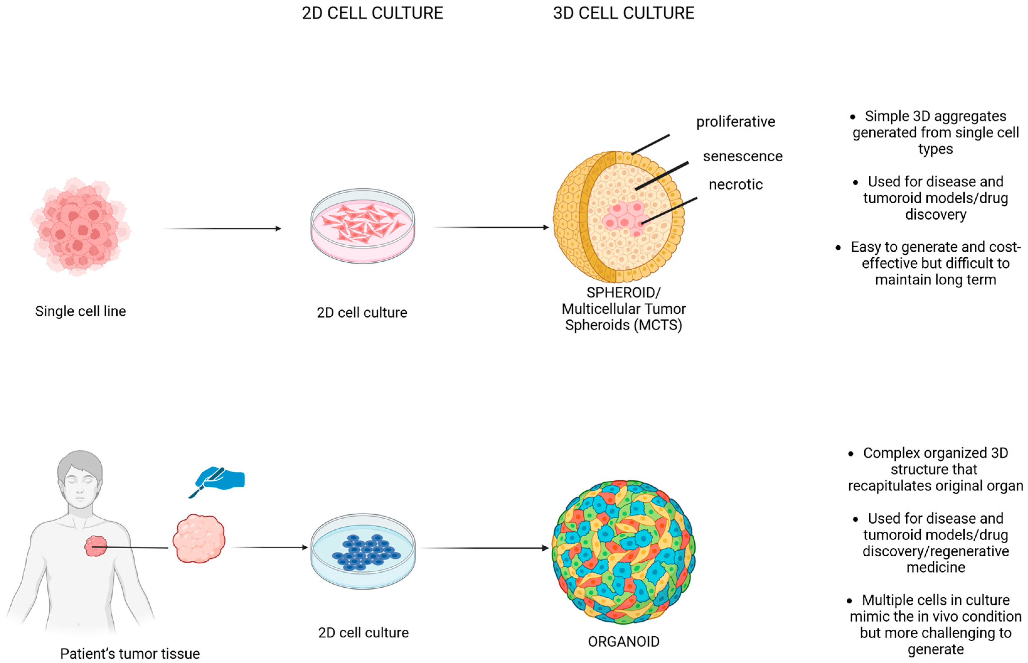
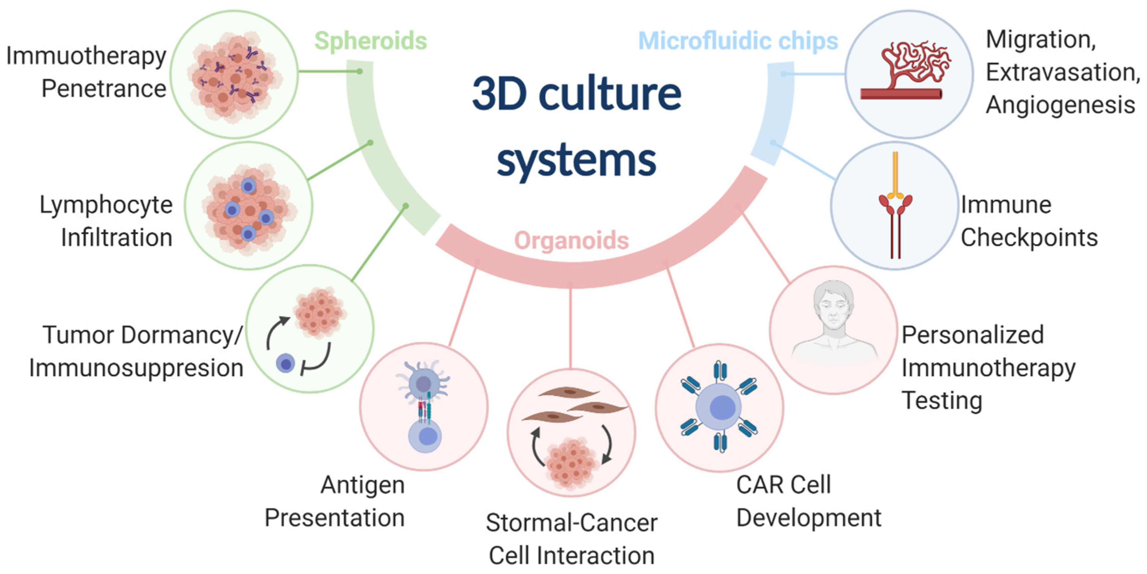
A 3D culture system is an advanced in vitro cellular growth environment that allows cells to proliferate, migrate, and interact in three dimensions, closely mimicking native extracellular matrix architecture and physiological tissue microenvironments compared to traditional two-dimensional monolayer cultures.
Introduction to 3D Cell Culture Systems
Cell biology research has historically relied on two-dimensional (2D) monolayer cultures grown on flat, rigid plastic or glass surfaces. While 2D cultures have provided foundational insights into cellular mechanisms, drug discovery, and molecular biology, they suffer from significant physiological limitations. Cells grown in a monolayer experience unnatural apical-basal polarization, uniform access to nutrients and oxygen, and a complete absence of the complex mechanical and biochemical cues provided by a natural extracellular matrix (ECM).
To bridge the gap between traditional in vitro assays and complex in vivo human physiology, researchers have turned to 3D culture systems. These advanced platforms recreate the multi-directional cell-cell and cell-matrix interactions that dictate native tissue architecture, gene expression, and cellular signaling. By establishing a physiologically relevant microenvironment, a 3D culture system significantly improves the predictive power of preclinical drug testing, cancer biology studies, regenerative medicine, and tissue engineering applications.
Understanding the landscape of 3D culture requires examining the underlying architectural types, material classifications, operational parameters, and integration methods that make these systems successful across diverse scientific disciplines.
Evolution from 2D Monolayers to 3D Environments
The transition from 2D to 3D culture is driven by the fundamental differences in how cells experience their surroundings. In a 2D environment, cells spread out on a rigid substrate, forming focal adhesions only on one plane. This mechanical constraint alters cell shape, nuclear architecture, and cytoskeletal tension, which in turn profoundly influences gene transcription and protein expression.
Conversely, a 3D culture system allows cells to assemble into complex multi-cellular structures—such as spheroids or organoids—where cells are completely surrounded by neighboring cells and ECM components. This multi-directional freedom restores physiological morphology, receptor distribution, and metabolic activity.
Biological and Mechanical Disparities
| Feature | 2D Monolayer Culture | 3D Culture System |
|---|---|---|
| Cellular Morphology | Flat, stretched, forced apical-basal polarity | Rounded, natural multi-axial spreading, in vivo-like architecture |
| Nutrient & Oxygen Gradient | Uniform exposure across all cells | Natural diffusion gradients (hypoxic core in large structures) |
| Cell-Matrix Interaction | Restricted to the bottom substrate interface | Dynamic, multi-directional engagement with surrounding ECM |
| Drug Sensitivity | Often artificially high due to uninhibited surface exposure | Realistic drug diffusion barriers, better mimicking clinical resistance |
| Gene & Protein Expression | Differs significantly from native tissues | Highly predictive of in vivo phenotypes and biomarker profiles |
Core Architecture and Classification of 3D Culture Systems
Three-dimensional culture platforms are generally divided into two primary categories based on their structural approach: scaffold-based systems and scaffold-free systems. Each category utilizes distinct physical and chemical mechanisms to support cellular aggregation and tissue formation.
Scaffold-Based 3D Systems
Scaffold-based platforms utilize artificial or natural structural matrices that act as a surrogate extracellular matrix. These matrices provide mechanical support, direct cellular organization, and facilitate the diffusion of nutrients and waste.
- Hydrogels: Composed of hydrophilic polymer networks that swell in water, hydrogels can be natural (e.g., collagen, fibrin, hyaluronic acid, Matrigel) or synthetic (e.g., polyethylene glycol, peptide amphiphiles). They offer tunable stiffness and biochemical signaling cues that mimic native soft tissues.
- Solid Scaffolds: Manufactured from porous ceramics, metals, or synthetic polymers (such as poly-lactic-co-glycolic acid), solid scaffolds offer high mechanical strength and are frequently utilized in bone tissue engineering and long-term structural implants.
- Nanofibrous Meshes: Electrospun or self-assembling peptide nanofibers create ultra-fine porous architectures that closely replicate the nanoscale fibrous structure of the natural ECM.
Scaffold-Free 3D Systems
Scaffold-free platforms encourage cells to aggregate spontaneously through cell-cell adhesion molecules (such as cadherins) without exogenous structural support.
- Spheroids and Organoids: Liquid-overlay techniques, hanging drop plates, and rotary bioreactors prevent cell adhesion to the culture vessel walls, forcing cells to coalesce into spherical aggregates or self-organizing organ-specific structures.
- Magnetic 3D Bioprinting: Utilizes biocompatible magnetic nanoparticles attached to cells, allowing external magnetic fields to rapidly assemble cells into desired spatial configurations.
Biomaterials and Biochemical Properties
The selection of biomaterials within a 3D culture system dictates its mechanical integrity, degradation rate, and biological compatibility. Researchers must balance biochemical complexity with experimental reproducibility.
Natural vs. Synthetic Polymers
Natural biomaterials possess inherent biological recognition sites, enzymatic degradability, and cellular remodeling capabilities. However, animal-derived products often exhibit batch-to-batch variability, undefined compositions, and potential immunogenicity. For instance, tumor-derived basement membrane extracts can contain variable growth factor profiles that complicate controlled experimentation.
Synthetic polymers, on the other hand, provide exquisite control over chemical composition, mechanical stiffness, and degradation kinetics. By functionalizing synthetic hydrogels with specific peptide sequences—such as adhesion ligands (RGD peptides)—scientists can engineer precisely customized microenvironments that isolate single variables without confounding background noise.
Mechanical Properties and Stiffness
Matrix stiffness is a critical mechanical cue that regulates stem cell differentiation, cancer metastasis, and cellular migration. Advanced 3D culture systems allow precise tuning of storage modulus ($G'$) and loss modulus ($G''$) to match the specific physiological elasticity of target tissues, ranging from soft neural tissue to rigid osteoid matrices.
Applications Across Scientific Disciplines
The adoption of 3D culture systems spans multiple advanced research domains, transforming how experimental models are designed and executed.
Preclinical Drug Discovery and Toxicology
Traditional high-throughput screening in 2D formats frequently yields high attrition rates during clinical trials because 2D models fail to predict human toxicity and efficacy accurately. 3D spheroids and organoids incorporate metabolic gradients, multi-layered cell resistance, and realistic drug penetration barriers. This enables pharmaceutical researchers to assess drug candidate cytotoxicity, penetration, and therapeutic windows with significantly higher translational fidelity.
Cancer Biology and Tumor Microenvironments
Tumors do not grow as monolayers; they exist within a complex, heterogeneous microenvironment characterized by hypoxia, dense extracellular matrices, stromal cells, and immune cell infiltration. A 3D tumor model allows researchers to study cancer stemness, epithelial-mesenchymal transition (EMT), angiogenesis, and immune evasion mechanisms in a context that mirrors in vivo solid tumors.
Regenerative Medicine and Tissue Engineering
In regenerative medicine, 3D culture systems serve as foundational frameworks for growing functional tissue constructs destined for transplantation. By seeding patient-derived stem cells onto customized scaffolds or bioinks, bioengineers can fabricate vascularized skin grafts, cardiac patches, cartilage constructs, and functional hepatic tissue.
Operational Workflow and Implementation Steps
Implementing a reliable 3D culture workflow requires careful planning, rigorous quality control, and adherence to specialized handling protocols.
- Experimental Design & Matrix Selection: Define whether the biological question requires a scaffold-based hydrogel or a scaffold-free spheroid model based on target tissue stiffness and imaging requirements.
- Cell Harvesting & Preparation: Expand cells in standard 2D culture until optimal passage numbers and viability (>90%) are achieved. Digest monolayers gently to maintain single-cell suspensions or small cell clusters.
- Seeding & Assembly: Introduce cells into the 3D platform (e.g., dispensing cell-hydrogel precursors into multi-well plates or distributing cell suspensions into ultra-low attachment plates for spheroid aggregation).
- Incubation & Gelation: For hydrogels, initiate crosslinking via thermal, ionic, or photo-initiated polymerization. Transfer cultures to humidified incubators maintained at standard physiological parameters ($37^\circ\text{C}$, 5% $\text{CO}_2$).
- Media Maintenance & Nutrient Exchange: Implement regular medium exchanges. Because 3D structures consume nutrients more rapidly and develop internal diffusion limitations, monitoring glucose consumption and pH shifts is vital.
- Harvesting & Analysis: Extract cells or whole constructs for downstream assays, utilizing specialized clearing agents for deep-tissue confocal imaging, enzymatic digestion for flow cytometry, or lysis protocols for molecular profiling.
Technical Challenges and Troubleshooting
Despite their vast advantages, working with 3D culture systems introduces unique operational and analytical hurdles.
Diffusion Limitations and Necrosis
In larger spheroids or dense hydrogels exceeding 200 to 400 micrometers in diameter, oxygen and nutrient diffusion become restricted. This leads to the spontaneous formation of a necrotic core surrounded by a hypoxic zone and an outer proliferating rim. While this mirrors solid tumor physiology, it can confound assays measuring bulk viability. Researchers must optimize initial seeding densities and monitor construct dimensions continuously.
Imaging and Optical Limitations
Thick 3D matrices scatter light, making high-resolution deep-tissue imaging difficult with standard brightfield or widefield microscopy. Advanced imaging requires multi-photon microscopy, confocal laser scanning microscopy, and optical tissue-clearing protocols that match the refractive index of the scaffold to preserve structural integrity while rendering the matrix transparent.
High-Throughput Scalability and Cost
Automating 3D culture workflows remains more challenging than conventional liquid handling for 2D plates. Hydrogel handling viscosity, precise pipetting requirements, and specialized multi-well plate configurations increase both material costs and technical complexity during high-throughput screening campaigns.
Frequently Asked Questions
What is the primary advantage of a 3D culture system over traditional 2D culture?
A 3D culture system provides a physiological microenvironment where cells experience multi-directional cell-cell and cell-matrix interactions, natural mechanical stiffness, and realistic nutrient gradients. This results in gene expression profiles and drug responses that closely mirror native in vivo tissues.
How do scaffold-free and scaffold-based 3D cultures differ?
Scaffold-free systems rely on spontaneous cell aggregation (such as spheroids or organoids) driven by endogenous cell-cell adhesion. Scaffold-based systems utilize natural or synthetic extracellular matrices—like hydrogels or porous polymers—to physically support, guide, and mechanically constrain cellular organization.
Why do large 3D spheroids develop a necrotic core?
As spheroids grow beyond a certain diameter (typically exceeding 200–400 micrometers), passive diffusion cannot supply sufficient oxygen and nutrients to the center of the structure, nor can it efficiently remove metabolic waste. This creates an inner hypoxic and necrotic zone similar to the microenvironment found in the interior of solid tumors.
Can standard analytical assays be used with 3D cultures?
Many standard assays (such as ATP luminescence, Western blotting, and flow cytometry) can be adapted for 3D cultures, but they frequently require specialized enzymatic digestion or physical disruption steps to release cells from the matrix. Furthermore, downstream imaging often requires optical clearing techniques to visualize deep structures accurately.
What factors dictate biomaterial selection for hydrogel-based systems?
Biomaterial selection is guided by required mechanical stiffness (elastic modulus), biocompatibility, degradation kinetics, cell adhesion ligand presentation, and transparency for optical imaging. Researchers must balance the biological realism of natural polymers with the precise reproducibility and tunability of synthetic polymers.
Conclusion and Recommendations
Adopting a 3D culture system represents a critical methodological upgrade for researchers seeking physiologically relevant in vitro models. To maximize success, laboratories should clearly define their biological endpoints before selecting a platform, ensuring that the chosen matrix stiffness, scale, and analysis pipeline align with experimental goals. By addressing diffusion limits, optimizing imaging protocols, and standardizing seeding densities, researchers can harness the full predictive power of three-dimensional cellular environments.