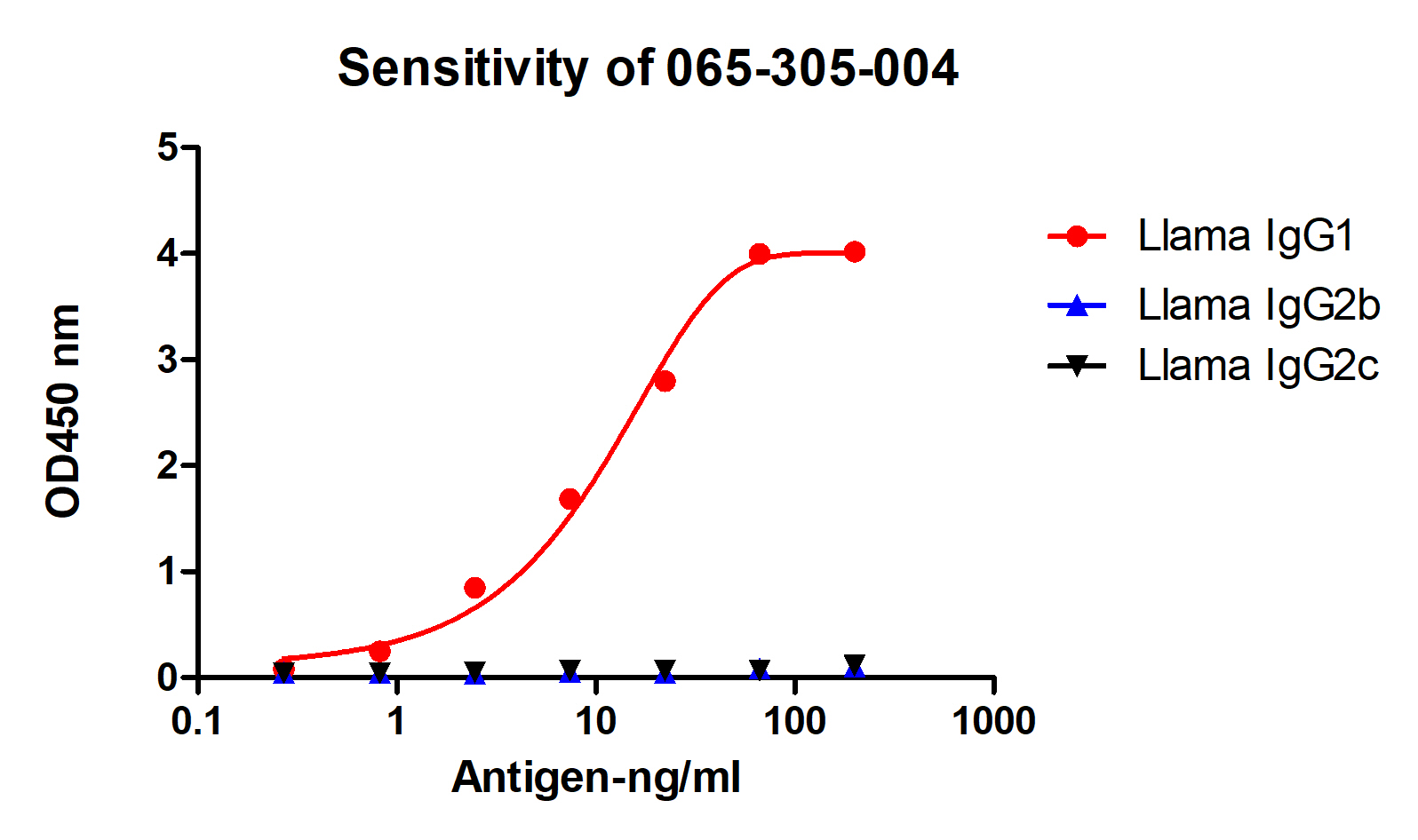
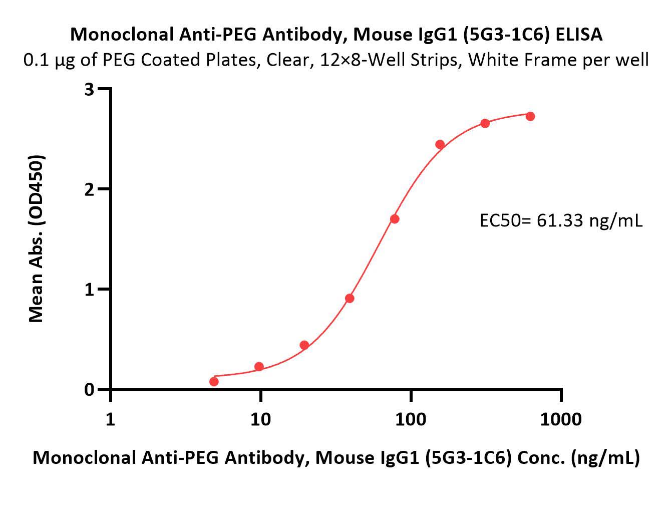
Anti-IgG1 antibodies are specialized binding proteins designed to target and interact with the immunoglobulin G subclass 1 (IgG1) isotype, playing a critical role in immunology, assay development, and targeted biotherapeutic monitoring. These reagents are essential tools for detecting, quantifying, and neutralizing human or murine IgG1 molecules in both diagnostic laboratories and advanced pharmacokinetic studies.
In the rapidly evolving landscape of modern biotechnology and clinical medicine, understanding the mechanics, applications, and limitations of anti-IgG1 reagents is vital for researchers and clinicians alike. Immunoglobulin G subclass 1 is the most abundant IgG subclass in human serum, making it a primary vehicle for therapeutic monoclonal antibodies. Consequently, anti-IgG1 antibodies serve as indispensable instruments for tracking drug clearance, evaluating immune responses, and ensuring the quality control of biologic drugs.
1. Structural Identity and Isotype Specificity of Anti-IgG1 Reagents
To understand how anti-IgG1 antibodies function, one must first examine the structural architecture of the immunoglobulin G molecule they target. Human IgG is divided into four distinct subclasses—IgG1, IgG2, IgG3, and IgG4—which share a high degree of sequence homology yet possess distinct functional properties, particularly within their constant heavy ($C_H$) domains and hinge regions. Anti-IgG1 antibodies are specifically engineered to recognize the unique antigenic determinants, or epitopes, located on the heavy chain of the IgG1 subclass.
The precision of these reagents relies on their ability to discriminate between IgG1 and closely related subclasses like IgG2 or IgG4. This specificity is achieved through rigorous hybridoma screening or recombinant expression techniques that select for clones binding exclusively to subclass-specific sequences, typically located in the $CH2$ or $CH3$ domains. Without this high degree of specificity, cross-reactivity would confound immunological assays, leading to false positives in multiplexing environments or inaccurate pharmacokinetic measurements.
Furthermore, anti-IgG1 antibodies can be generated as monoclonal or polyclonal preparations, each serving different experimental needs. Monoclonal anti-IgG1 antibodies offer exceptional batch-to-batch reproducibility and precise epitope mapping, making them ideal for quantitative ELISA (Enzyme-Linked Immunosorbent Assay) development. Polyclonal anti-IgG1 antibodies, harvested from immunized host species such as goats or rabbits, recognize multiple epitopes across the IgG1 constant region, providing robust capture capabilities in complex biological matrices where target conformation might be partially altered.
2. Critical Applications in Therapeutic Monoclonal Antibody Monitoring
The advent of monoclonal antibody (mAb) therapies has revolutionized the treatment of oncology, autoimmune disorders, and infectious diseases. Because a vast majority of these therapeutic mAbs—such as trastuzumab, rituximab, and pembrolizumab—belong to the human IgG1 isotype due to their robust effector functions, anti-IgG1 antibodies have become foundational components of therapeutic drug monitoring (TDM) and pharmacokinetic (PK) profiling.
During clinical trials and post-marketing surveillance, researchers must accurately measure the concentration of the therapeutic antibody in patient serum over time. Anti-IgG1 reagents are frequently employed as capture or detection antibodies in bridging ELISAs or liquid chromatography-tandem mass spectrometry (LC-MS/MS) workflows. These assays enable scientists to determine drug clearance rates, half-life, and bioavailability, ensuring that patients receive therapeutic doses within the optimal window of efficacy and safety.
| Application Field | Primary Function of Anti-IgG1 | Key Assay Format |
|---|---|---|
| Pharmacokinetics (PK) | Quantifying therapeutic IgG1 drug levels in serum | Bridging ELISA / Gyrolab |
| Immunogenicity Testing | Detecting Anti-Drug Antibodies (ADAs) | Electrochemiluminescence (ECL) |
| Flow Cytometry | Staining cell-surface bound human IgG1 targets | Direct/Indirect Immunofluorescence |
| Quality Control | Verifying structural integrity during manufacturing | Western Blot / SEC-HPLC |
Beyond standard pharmacokinetic tracking, anti-IgG1 antibodies are instrumental in anti-drug antibody (ADA) assays. When patients receive biologic drugs, their immune systems may mount an adaptive response, producing anti-drug antibodies that can neutralize the therapeutic agent or accelerate its clearance. Specialized anti-IgG1 reagents help characterize the immune response, distinguishing between pre-existing immunoglobulins and treatment-induced antibodies.
3. Mechanisms of Effector Function Modulation and Neutralization
Beyond mere detection, anti-IgG1 antibodies are frequently utilized to manipulate or study the biological effector functions inherent to the IgG1 isotype. Human IgG1 is renowned for its potent engagement with the innate immune system, mediated primarily through two major pathways: antibody-dependent cellular cytotoxicity (ADCC) and complement-dependent cytotoxicity (CDC). These mechanisms depend on the interaction between the Fc (fragment crystallizable) region of the IgG1 antibody and cellular Fc receptors (such as Fc$\gamma$RIIIa) or the C1q component of the complement cascade.
In experimental settings, researchers use anti-IgG1 antibodies—or engineered fragments thereof, such as Fab or scFv formats—to block these Fc-mediated interactions. By masking specific residues on the IgG1 Fc region, investigators can dissect whether a therapeutic antibody's clinical effect stems from direct target neutralization or immune-mediated cell lysis. Conversely, anti-IgG1 cross-linking agents can be deployed to artificially cluster surface receptors in vitro, mimicking the multivalent binding of immune complexes to trigger downstream signaling pathways in cellular immunology studies.
It is also important to note the distinction between anti-IgG1 reagents used as analytical tools and therapeutic anti-IgG1 molecules themselves. While research reagents are designed to bind and detect IgG1 molecules, therapeutic anti-IgG antibodies (such as those targeting specific disease-associated immunoglobulins in autoimmune diseases) act as active biological drugs designed to deplete pathological B-cell clones or neutralize aberrant immune complexes. Understanding this duality prevents confusion between diagnostic assay components and targeted immunotherapy agents.
4. Technical Challenges and Cross-Reactivity Considerations in Laboratory Assays
Deploying anti-IgG1 antibodies in complex biological environments presents several distinct biochemical hurdles. The primary challenge is mitigating cross-reactivity with endogenous immunoglobulins. Because human serum contains massive quantities of polyclonal IgG distributed across all four subclasses (with IgG1 typically making up 60% to 70% of total serum IgG), analytical assays are constantly prone to background noise and matrix interference.
To overcome these limitations, assay developers must optimize blocking buffers and employ stringent washing protocols. When designing a sandwich ELISA, for instance, the anti-IgG1 capture antibody must exhibit negligible affinity for IgG2, IgG3, and IgG4 to prevent false elevation of signal driven by non-target immunoglobulins. Furthermore, rheumatoid factor (RF)—an autoantibody directed against the Fc region of human IgG—frequently causes false-positive results in clinical immunoassays by bridging capture and detection antibodies independently of the target analyte.
| Challenge Factor | Underlying Cause | Mitigation Strategy |
|---|---|---|
| Subclass Cross-Reactivity | High structural homology across IgG1-4 | Use affinity-purified monoclonal reagents with mapped epitopes |
| Matrix Interference | High abundance of endogenous serum proteins | Implement optimized blocking buffers and sample dilution |
| Rheumatoid Factor (RF) | Autoantibodies binding human Fc regions | Add blocking reagents or use Fab'2 assay fragments |
| HAMA/HAHA Effects | Human anti-mouse/animal antibodies in patient samples | Incorporate isotype-matched animal serum controls |
Another critical factor is species cross-reactivity. Anti-human IgG1 antibodies are generally optimized for human samples and may fail to recognize murine, simian, or canine IgG1 subclasses due to evolutionary divergence in the constant region sequences. Researchers conducting translational studies in non-human primate models must carefully select anti-IgG reagents validated specifically for cross-species reactivity, ensuring that preclinical pharmacokinetic data accurately reflects expected human dynamics.
5. Regulatory Standards and Quality Control in Reagent Manufacturing
The reliability of diagnostic and therapeutic research depends heavily on the rigorous quality control of anti-IgG1 antibodies. Because these reagents are utilized in regulated environments—such as Good Laboratory Practice (GLP) preclinical trials and Good Clinical Practice (GCP) biomarker analysis—manufacturers must adhere to strict validation frameworks to ensure lot-to-lot consistency, stability, and high affinity.
Quality assurance protocols for commercial anti-IgG1 antibodies typically involve advanced analytical techniques such as Surface Plasmon Resonance (SPR) to measure association and dissociation rate constants ($k_a$ and $k_d$), thereby verifying binding affinity. Additionally, purity is evaluated using Size-Exclusion Chromatography (SEC-HPLC) and capillary electrophoresis to detect protein aggregation, fragmentation, or contamination by carrier proteins such as bovine serum albumin (BSA).
Regulatory bodies also emphasize the importance of comprehensive documentation, including detailed datasheets specifying recommended dilutions, tested assay platforms, and known cross-reactivity profiles. Researchers selecting anti-IgG1 reagents for quantitative work must verify that the supplier provides validated performance data for the specific intended application, whether that involves Western blotting, immunohistochemistry, or high-throughput clinical screening platforms.
Frequently Asked Questions About Anti-IgG1 Antibodies
What is the primary function of an anti-IgG1 antibody in research?
Anti-IgG1 antibodies are primarily used to specifically detect, capture, or quantify immunoglobulin G subclass 1 molecules. In research and clinical settings, they enable the measurement of therapeutic monoclonal antibody concentrations, support immunogenicity testing, and help characterize immune responses by distinguishing IgG1 from other immunoglobulin isotypes.
Can anti-IgG1 antibodies cross-react with other IgG subclasses?
High-quality anti-IgG1 antibodies are engineered to minimize cross-reactivity with IgG2, IgG3, and IgG4 subclasses. However, lower-affinity or poorly characterized polyclonal preparations may exhibit varying degrees of cross-reactivity due to the high structural sequence homology shared across human IgG heavy chains.
Are anti-IgG1 reagents used in clinical treatments or diagnostic assays?
While some therapeutic antibodies are targeted against pathological immunoglobulins, the vast majority of commercial "anti-IgG1" products are analytical research reagents used in laboratory assays such as ELISAs, Western blots, and flow cytometry. They serve as tools to monitor drug levels rather than acting as primary therapeutic drugs themselves.
How do researchers prevent matrix interference when using anti-IgG1 in serum assays?
Matrix interference from endogenous serum proteins and rheumatoid factor is mitigated by using highly specific monoclonal capture reagents, incorporating optimized blocking agents (such as bovine serum albumin or non-fat dry milk), and applying appropriate sample dilution curves to maintain assay linearity and specificity.
What factors should be considered when selecting an anti-IgG1 antibody for pharmacokinetic studies?
Researchers must evaluate target species specificity, affinity constants, format suitability (monoclonal vs. polyclonal), and validation status for specific analytical platforms such as Gyrolab, Meso Scale Discovery (MSD) electrochemiluminescence, or standard colorimetric ELISAs.
To successfully integrate anti-IgG1 reagents into your analytical workflow, begin by mapping the precise structural requirements of your assay platform, verifying subclass specificity against potential endogenous interferents, and selecting affinity-validated monoclonal options backed by robust batch-specific quality control data.