Ascitic fluid interpretation is the systematic clinical and laboratory analysis of fluid collected from the peritoneal cavity to determine the underlying cause of ascites. By utilizing the Serum-Ascites Albumin Gradient (SAAG) alongside cell counts, total protein, and specialized markers, clinicians can accurately differentiate between portal hypertension, malignancy, infection, and rare inflammatory conditions.
Ascites, the pathological accumulation of fluid within the peritoneal cavity, is a clinical manifestation of various systemic and localized diseases. While cirrhosis remains the most common cause in the developed world, accounting for approximately 80% of cases, other etiologies such as malignancy, heart failure, and tuberculosis must be rigorously excluded. In 2026, the standard of care for any patient with new-onset ascites or a significant change in clinical status remains a diagnostic paracentesis followed by a structured interpretation of the ascitic fluid.
Medical Disclaimer: This article is intended for educational purposes for healthcare professionals. Clinical decisions should be based on individual patient presentation and current institutional guidelines. All laboratory values must be interpreted within the specific clinical context.
The Role of Paracentesis in Diagnostic Evaluation
Diagnostic paracentesis is the gold standard for evaluating peritoneal fluid. It is a relatively safe bedside procedure with a low risk of complications, even in patients with coagulopathy. The primary goal of fluid analysis is to categorize the fluid based on its biochemical and cellular properties, which narrows the differential diagnosis significantly.
Paracentesis is indicated for:
- New-onset ascites: To establish the primary diagnosis.
- Hospital admission: All patients with cirrhosis and ascites admitted to the hospital should undergo paracentesis to rule out Spontaneous Bacterial Peritonitis (SBP), as symptoms can be subtle or absent.
- Clinical deterioration: Fever, abdominal pain, worsening renal function, or hepatic encephalopathy in a patient with known ascites.
Once the fluid is obtained, it should be sent immediately to the laboratory. For the most accurate results, ascitic fluid should be inoculated into blood culture bottles at the bedside, as this significantly increases the sensitivity for detecting bacterial growth compared to traditional laboratory plating.
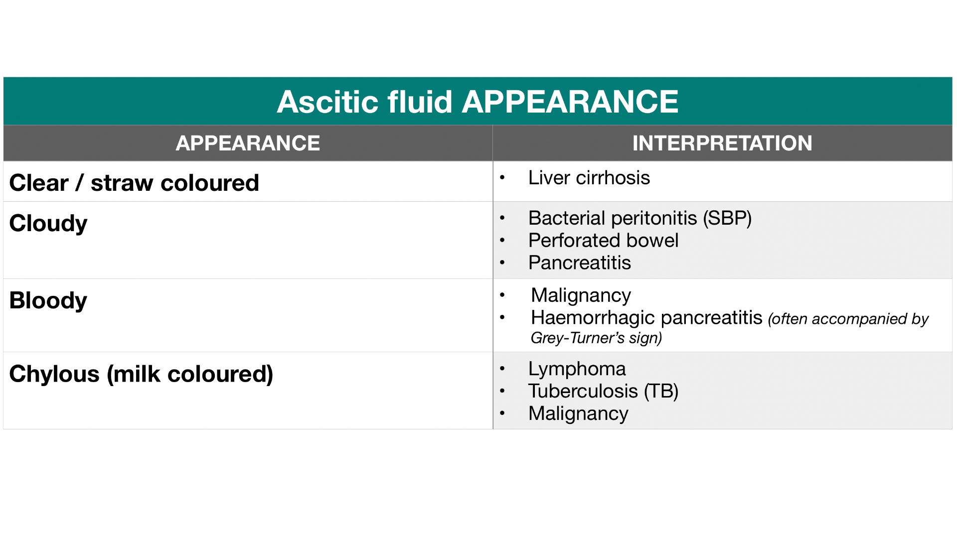
Gross Physical Appearance of Ascitic Fluid
The initial visual assessment of the fluid provides immediate, though non-specific, diagnostic clues.
- Normal/Cirrhotic: Typically clear or straw-colored.
- Cloudy/Turbid: Suggests an elevated white blood cell count, often indicative of infection (SBP) or occasionally malignancy.
- Bloody: Often a result of a "traumatic tap" during the procedure. However, if the fluid is non-clearing or homogeneously bloody, it may suggest malignancy (especially hepatocellular carcinoma or peritoneal carcinomatosis) or a ruptured ectopic pregnancy. If the fluid is bloody, the neutrophil count must be adjusted (subtract 1 PMN for every 250 red blood cells).
- Milky (Chylous): Indicates the presence of thoracic duct obstruction or damage, resulting in the leakage of lymph rich in triglycerides. Common causes include lymphoma or post-surgical complications.
- Brown/Green: May indicate a high bilirubin concentration, often seen in gallbladder perforation or perforated duodenal ulcers.
The Serum-Ascites Albumin Gradient (SAAG)
The SAAG has replaced the traditional "transudate vs. exudate" classification, which was found to be unreliable in the peritoneal cavity. The SAAG is calculated by subtracting the albumin concentration of the ascitic fluid from the albumin concentration of a serum sample obtained on the same day.
Formula: SAAG = Serum Albumin - Ascitic Fluid Albumin
The SAAG concept is based on Starling’s forces. Portal hypertension increases the hydrostatic pressure in the hepatic sinusoids, which "pushes" fluid into the peritoneum. However, the oncotic pressure (maintained by albumin) resists this. A high gradient indicates that the pressure difference is significant, usually due to portal hypertension.
High Gradient (SAAG ≥ 1.1 g/dL)
A SAAG of 1.1 g/dL or greater indicates the presence of portal hypertension with approximately 97% accuracy. Common causes include:
- Cirrhosis: The most frequent cause.
- Alcoholic Hepatitis: Often presents with very high SAAG and high protein.
- Congestive Heart Failure: Characterized by a high SAAG and a total protein level > 2.5 g/dL.
- Budd-Chiari Syndrome: Acute hepatic vein thrombosis.
- Massive Hepatic Metastases: Replacing significant liver parenchyma.
- Portal Vein Thrombosis: Isolated portal hypertension.
Low Gradient (SAAG < 1.1 g/dL)
A SAAG of less than 1.1 g/dL indicates that portal hypertension is not the driver of fluid accumulation. These cases are usually related to inflammation, increased capillary permeability, or lymphatic leakage. Causes include:
- Peritoneal Carcinomatosis: Primary or metastatic peritoneal tumors.
- Tuberculous Peritonitis: Often presents with a high lymphocyte count.
- Pancreatitis: Usually associated with very high ascitic amylase levels.
- Nephrotic Syndrome: Low serum albumin leads to fluid leakage.
- Serositis: Seen in systemic lupus erythematosus or other connective tissue diseases.
Cellular Analysis and Differential Count
The cell count is the most critical test for excluding or confirming infection. The total white blood cell (WBC) count and the absolute polymorphonuclear neutrophil (PMN) count must be calculated.
Spontaneous Bacterial Peritonitis (SBP)
SBP is defined as an ascitic fluid PMN count ≥ 250 cells/mm³ in the absence of an intra-abdominal, surgically treatable source of infection. Even if the culture is negative (culture-negative neutrocytic ascites), the patient must be treated with antibiotics if the PMN count exceeds this threshold.
Secondary Bacterial Peritonitis
It is vital to distinguish SBP from secondary peritonitis (caused by a perforated viscus or abscess). Secondary peritonitis is suggested if the PMN count is very high (> 1,000 cells/mm³) or if at least two of the following "Runyon's Criteria" are met:
- Total protein > 1 g/dL.
- Glucose < 50 mg/dL.
- LDH greater than the upper limit of normal for serum.
Lymphocytic Ascites
An elevated total WBC count with a lymphocyte predominance (> 50-70%) is suspicious for:
- Tuberculosis.
- Peritoneal Carcinomatosis.
- Lymphoma.
Total Protein and its Clinical Significance
While SAAG determines the presence of portal hypertension, the total protein concentration helps further narrow the etiology within those categories.
- High Protein Ascites (> 2.5 g/dL): Seen in heart failure, Budd-Chiari syndrome, and early-stage peritoneal malignancy. In heart failure, the liver sinusoids are congested but the "pores" are large enough to allow some protein through, leading to a high SAAG and high protein.
- Low Protein Ascites (< 2.5 g/dL): Typically seen in cirrhosis and nephrotic syndrome. Patients with cirrhosis and very low ascitic protein (< 1.5 g/dL) are at an increased risk of developing SBP and may require prophylactic antibiotics.
Secondary and Specialized Diagnostic Tests
In cases where the diagnosis remains ambiguous after the initial SAAG and cell count, specialized tests are warranted.
Ascitic Amylase
If pancreatic ascites (due to pancreatitis or a pancreatic duct leak) is suspected, ascitic amylase should be measured. In these cases, the fluid amylase level is typically much higher than the serum amylase level (often > 1,000 U/L).
Ascitic Glucose and LDH
As mentioned in Runyon's criteria, low glucose and high LDH are markers of significant inflammation or bacterial consumption. In SBP, glucose is usually similar to serum levels, whereas in secondary peritonitis, it drops significantly.
Cytology
Cytology is used to detect malignant cells. While highly specific, it only has a sensitivity of about 60-70% for peritoneal carcinomatosis and is often negative in cases of primary hepatocellular carcinoma because the tumor rarely seeds the peritoneum.
Adenosine Deaminase (ADA)
ADA is a useful marker for tuberculous peritonitis, particularly in high-prevalence areas. A high ADA (typically > 30-40 U/L) is highly suggestive of TB, though it may be falsely elevated in some malignancies.
Triglycerides
Levels > 200 mg/dL (and usually much higher) confirm a diagnosis of chylous ascites.
Summary Table of Ascitic Fluid Interpretation
| Etiology | SAAG | Total Protein | PMN Count | Other Findings |
|---|---|---|---|---|
| Cirrhosis | ≥ 1.1 g/dL | < 2.5 g/dL | < 250/mm³ | Low glucose common in advanced stages |
| Cardiac Ascites | ≥ 1.1 g/dL | > 2.5 g/dL | < 250/mm³ | High serum BNP levels |
| SBP | ≥ 1.1 g/dL | < 2.5 g/dL | ≥ 250/mm³ | Culture positive in 50-70% |
| Peritoneal Carcinomatosis | < 1.1 g/dL | > 2.5 g/dL | Variable | Positive cytology (60-70%) |
| Tuberculous Peritonitis | < 1.1 g/dL | > 2.5 g/dL | > 250/mm³ | Lymphocyte predominance; High ADA |
| Pancreatic Ascites | < 1.1 g/dL | > 2.5 g/dL | Variable | Amylase > 1,000 U/L |
| Nephrotic Syndrome | < 1.1 g/dL | < 2.5 g/dL | < 250/mm³ | Proteinuria on urinalysis |
Common Pitfalls and Troubleshooting
Interpreting ascitic fluid is generally straightforward, but several scenarios can complicate the clinical picture:
- The "Bloody Tap": If the needle hits a vessel during paracentesis, the RBC and WBC counts will be artificially elevated. To correct the PMN count, subtract 1 PMN for every 250 RBCs measured in the fluid. If the corrected PMN count is still ≥ 250, treat for SBP.
- Recent Diuresis: Vigorous treatment with diuretics can increase the concentration of protein and the SAAG score slightly. However, it rarely moves a patient from one SAAG category to another.
- Hypoalbuminemia: If the serum albumin is very low (< 1.1 g/dL), the SAAG may be falsely low because the gradient cannot mathematically reach the 1.1 threshold. In these rare cases, clinicians must rely more heavily on total protein and clinical context.
- Mixed Ascites: A patient may have two causes of ascites simultaneously (e.g., cirrhosis and peritoneal carcinomatosis). This often results in a "High SAAG" (driven by portal hypertension) but with an unexpectedly high protein or positive cytology.
Clinical Workflow for Fluid Interpretation
To ensure no diagnostic data is missed, a standardized workflow is recommended:
- Immediate Bedside Action: Assess color and clarity. Inoculate blood culture bottles immediately.
- Laboratory Orders: Order SAAG (Albumin in both serum and fluid), Cell Count with differential, and Total Protein.
- Triggered Orders: If fluid is cloudy, ensure LDH and Glucose are prioritized. If the patient has a history of malignancy, order Cytology. If there is a history of pancreatitis or recent abdominal trauma, order Amylase.
- Verification: Compare the SAAG to the protein level. A high SAAG/Low Protein combination is the classic signature of cirrhosis.
- Intervention: If PMNs are ≥ 250, initiate empiric antibiotics (e.g., a third-generation cephalosporin) regardless of the patient's temperature or white blood cell count in the blood.
Frequently Asked Questions
Can you have SBP with a SAAG less than 1.1?
While theoretically possible if a patient has a non-portal hypertensive cause of ascites (like malignancy) that becomes secondarily infected, the vast majority of SBP cases occur in the setting of portal hypertension (SAAG ≥ 1.1). If a low SAAG fluid has high PMNs, clinicians should look aggressively for a surgical cause (secondary peritonitis).
Why do we use SAAG instead of the exudate/transudate model?
The exudate/transudate model, based on total protein, was originally designed for pleural effusions. In the peritoneum, it incorrectly classified up to 20-30% of patients. Many patients with cirrhosis have high-protein fluid, and many with malignancy have low-protein fluid. The SAAG is much more accurate (97%) because it directly reflects the pathophysiology of portal hypertension.
Is a culture necessary if the PMN count is high?
Yes. While treatment should start immediately based on the PMN count, the culture is vital for identifying the specific organism and its antibiotic sensitivities. This allows for narrowing the antibiotic spectrum once the results are available, typically after 48-72 hours.
What should I do if the ascitic fluid is milky?
A milky appearance indicates chylous ascites. You should order an ascitic triglyceride level. If the level is > 200 mg/dL, the diagnosis is confirmed. The next steps usually involve imaging (CT or lymphangiography) to look for lymphoma, lymphatic obstruction, or evidence of surgical injury to the lymphatic system.
How often should paracentesis be repeated?
In patients being treated for SBP, a repeat paracentesis is not always necessary if the patient is showing clear clinical improvement. However, if there is no improvement or if the clinical condition worsens after 48 hours of antibiotics, a repeat tap is mandatory to ensure the PMN count is decreasing (it should drop by at least 25% from the baseline).
Final Recommendation
When interpreting ascitic fluid, always prioritize the SAAG to determine if portal hypertension is present, followed immediately by the PMN count to rule out SBP. Never wait for culture results to begin antibiotics if the PMN count is ≥ 250 cells/mm³. For cases that do not fit the classic "High SAAG, Low Protein" profile of cirrhosis, expand your search to include malignancy, heart failure, and rare infections using protein, cytology, and amylase markers.