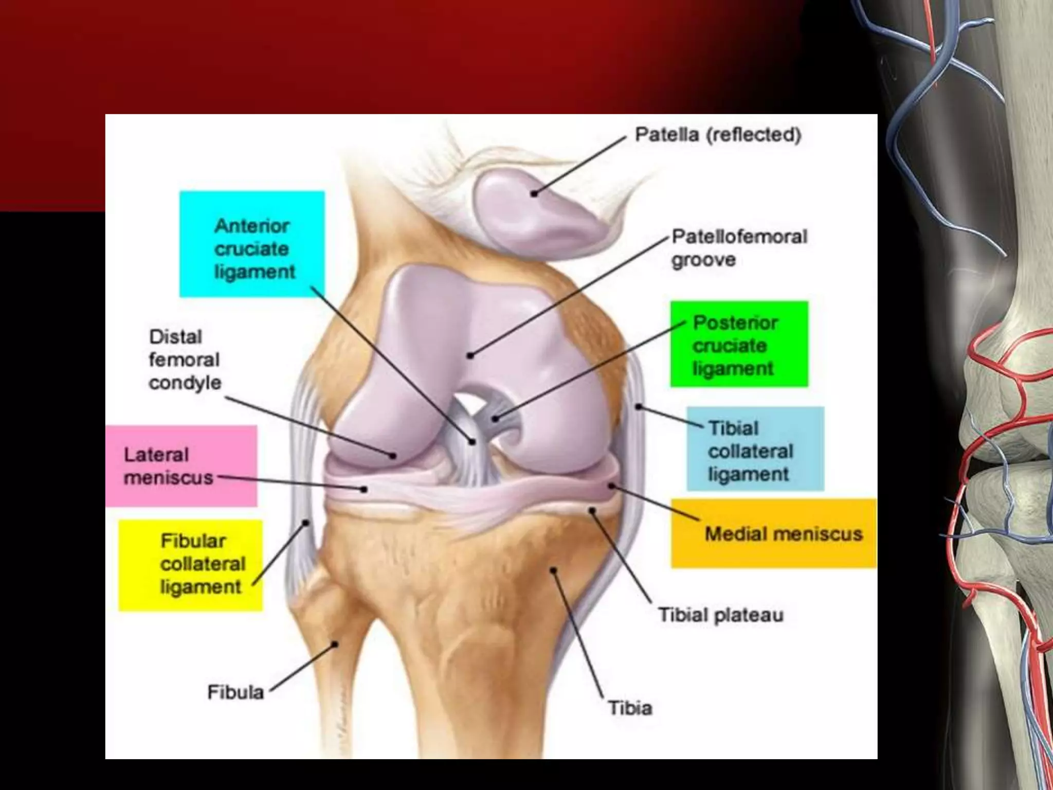
The human knee joint consists of three distinct anatomical compartments enclosed within a single, continuous synovial capsule: the medial tibiofemoral compartment, the lateral tibiofemoral compartment, and the patellofemoral compartment. Together, these compartments distribute mechanical loads, enable fluid flexion and extension, and maintain lower extremity stability during weight-bearing activities.
Disclaimer: This article is intended strictly for educational and informational purposes and does not constitute formal medical advice, diagnosis, or treatment. Always consult a qualified orthopedic specialist or healthcare provider regarding knee pain, structural injuries, or surgical planning.
Understanding the Tri-Compartmental Architecture of the Knee
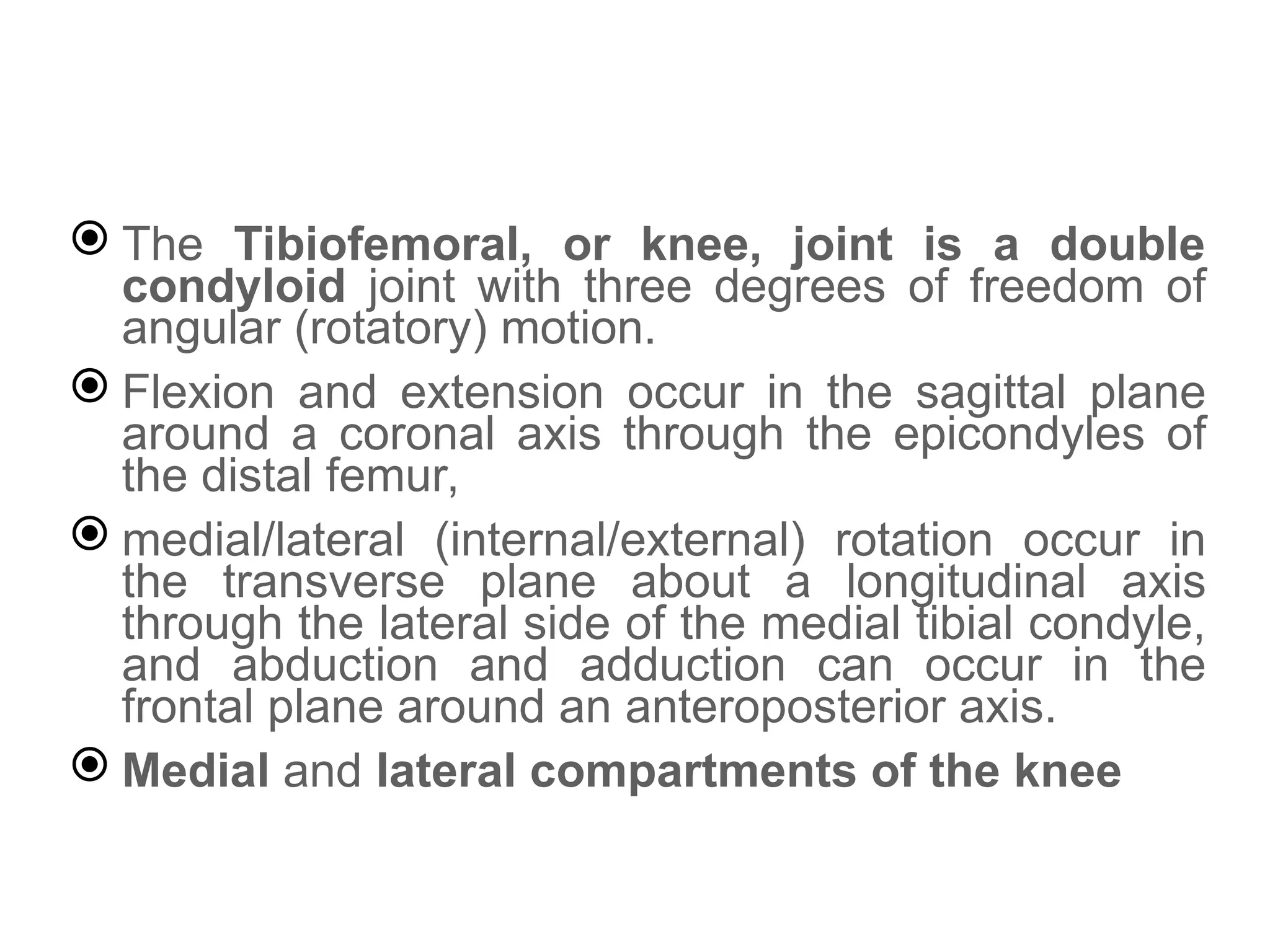
Although the knee is frequently referred to as a simple hinge joint (ginglymus), it is functionally a complex, modified hinge joint (trochoginglymus). It permits not only axial flexion and extension, but also a small degree of internal and external rotation during movement.
The structural foundation of the knee rests upon the articulation of three major bones: the distal femur (thigh bone), the proximal tibia (shin bone), and the patella (kneecap). Rather than operating as a single, uniform articular chamber, the knee joint space is compartmentalized into three primary functional zones.
Understanding the individual boundaries and mechanics of these three compartments is vital for diagnosing joint pathologies, understanding load distribution during gait, and guiding interventions ranging from targeted physical therapy to partial or total knee arthroplasty.
- [ Femur ]
- Patellofemoral Compartment (Anterior)
- Medial Tibiofemoral Compartment (Inner)
- Lateral Tibiofemoral Compartment (Outer)
- [ Tibia ]
The Continuous Synovial Cavity and Capsular Constraints
While clinicians describe the knee as having three discrete compartments, it is essential to note that these areas share a continuous synovial cavity. A fibrous joint capsule lined with a specialized synovial membrane encloses the entire joint space, secreting synovial fluid that lubricates the hyaline cartilage surfaces and supplies oxygen and nutrients to avascular articular tissues.
The internal division into distinct compartments arises from the anatomical geometry of the femoral condyles, the corresponding tibial plateaus, and the anterior trochlear groove. Ligamentous structures, meniscal tissue, and synovial folds (plica) further delineate these regions mechanically and functionally. When injury or disease affects one area, the shared synovial cavity means inflammatory responses can spread throughout the joint, even while structural damage remains confined to a single compartment.
The Medial Tibiofemoral Compartment
The medial tibiofemoral compartment occupies the inner aspect of the knee joint. It forms the articulation between the rounded medial femoral condyle and the relatively concave medial tibial plateau.
| Feature | Medial Tibiofemoral Compartment |
|---|---|
| Articulating Surfaces | Medial Femoral Condyle & Medial Tibial Plateau |
| Primary Meniscus | C-Shaped Medial Meniscus (Less Mobile, Anchored) |
| Primary Ligament | Medial Collateral Ligament (MCL) |
| Weight-Bearing Burden | ~60% to 70% of static/gait axial load |
| Primary Malalignment | Varus Deformity ("Bow-legged") |
Structural Components of the Medial Compartment
- Medial Femoral Condyle: Larger, longer, and more curved than its lateral counterpart, providing a broad surface for weight distribution.
- Medial Tibial Plateau: Slightly concave in structure, offering a stable socket-like receiver for the medial femoral condyle.
- Medial Meniscus: A semi-lunar, C-shaped fibrocartilaginous structure attached firmly around its periphery to the joint capsule and the deep fibers of the medial collateral ligament.
- Medial Collateral Ligament (MCL): A broad, flat band providing primary resistance against valgus (inward collapse) stress.
Because the medial meniscus is securely anchored to the joint capsule and the deep layer of the MCL, its mobility is significantly limited compared to the lateral meniscus. This anatomical rigidity makes the medial meniscus particularly vulnerable to shearing forces during twisting or pivoting movements.
Biomechanics and Susceptibility to Wear
In a neutrally aligned lower extremity, the mechanical axis of the leg (the line drawn from the center of the femoral head to the center of the ankle joint) passes slightly medial to the center of the knee. As a result, the medial tibiofemoral compartment carries approximately 60% to 70% of the net weight-bearing load during normal standing and walking.
This disproportionate load allocation makes the medial compartment the most common site for primary osteoarthritis (unicompartmental osteoarthritis). As cartilage wears down inside the medial compartment, loss of joint space height can lead to progressive varus alignment (a "bow-legged" posture). This alignment shift shifts the mechanical axis further inward, creating a dynamic feedback loop that accelerates wear on the medial articular cartilage and meniscus.
The Lateral Tibiofemoral Compartment
The lateral tibiofemoral compartment is located on the outer side of the knee joint. It is formed by the articulation between the lateral femoral condyle and the lateral tibial plateau.
| Feature | Lateral Tibiofemoral Compartment |
|---|---|
| Articulating Surfaces | Lateral Femoral Condyle & Lateral Tibial Plateau |
| Primary Meniscus | O-Shaped Lateral Meniscus (Highly Mobile) |
| Primary Ligament | Lateral Collateral Ligament (LCL) |
| Weight-Bearing Burden | ~30% to 40% of static axial load |
| Primary Malalignment | Valgus Deformity ("Knock-kneed") |
Structural Features and Meniscal Mobility
- Lateral Femoral Condyle: Shorter and broader anteroposteriorly than the medial condyle, with a prominent anterior wall that helps prevent lateral subluxation of the patella.
- Lateral Tibial Plateau: Slightly convex anteroposteriorly, creating an inherently less stable "ball-on-ball" resting geometry compared to the medial side.
- Lateral Meniscus: Almost circular (O-shaped), covering a larger surface area of the tibial plateau than the medial meniscus. Critically, it is not attached to the lateral collateral ligament and has fewer capsular attachments.
- Lateral Collateral Ligament (LCL): A cord-like structure that originates on the lateral femoral epicondyle and inserts onto the head of the fibula, functioning independently of the joint capsule.
The loose capsular attachments of the lateral meniscus allow it to slide anteroposteriorly by up to 10–12 millimeters during joint flexion. This increased mobility helps accommodate the complex rotation of the lateral femoral condyle during deep knee bending and reduces the risk of dynamic entrapment or shearing tears compared to the medial meniscus.
Biomechanical Distinctions from the Medial Side
Because the lateral tibial plateau is convex, joint stability in the lateral compartment relies heavily on surrounding soft tissues, including the popliteus tendon, the iliotibial (IT) band, and the lateral meniscal roots.
When structural failure or cartilage loss occurs in this compartment, it often leads to or results from a valgus alignment ("knock-kneed" stance). Valgus alignment shifts the mechanical load vector laterally, concentrating forces across the lateral femoral condyle and lateral tibial plateau.
Although lateral unicompartmental osteoarthritis is less frequent than medial disease, it presents distinct surgical challenges due to dynamic changes in soft-tissue tension along the lateral side of the knee.
The Patellofemoral Compartment
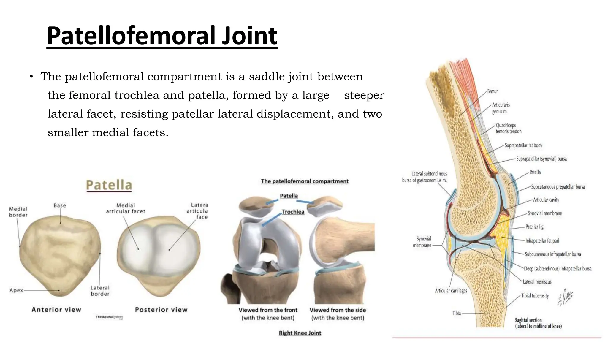
The patellofemoral compartment is situated in the anterior (front) region of the knee joint. It encompasses the posterior articular surface of the patella and the trochlear groove (trochlea) of the anterior distal femur.
| Feature | Patellofemoral Compartment |
|---|---|
| Articulating Surfaces | Posterior Patella & Femoral Trochlear Groove |
| Thickest Cartilage | Patellar Cartilage (up to 5-6 mm thick) |
| Driving Force | Extensor Mechanism (Quadriceps & Patellar Tendon) |
| Key Stabilizer | Medial Patellofemoral Ligament (MPFL) |
| Primary Pathologies | Patellofemoral Pain Syndrome, Instability |
The Anterior Articular Mechanism
The patella is the largest sesamoid bone in the human body, embedded within the quadriceps tendon superiorly and anchored to the tibial tuberosity inferiorly via the patellar tendon (patellar ligament).
- Posterior Patellar Articular Surface: Divided by a vertical ridge into a larger lateral facet and a smaller medial facet (along with an occasional odd facet on the most medial edge).
- Trochlear Groove: A V-shaped depression on the anterior femur designed to guide the patella as the knee flexes and extends.
- Articular Cartilage Depth: The hyaline cartilage on the posterior surface of the patella is among the thickest in the human body, measuring up to 5 to 6 millimeters to withstand compressive forces.
Patellar Tracking Dynamics and Biomechanical Load
The patella acts as a biomechanical fulcrum. By increasing the distance (moment arm) between the quadriceps tendon and the joint's axis of rotation, it increases the mechanical advantage of the quadriceps muscle group by up to 30% to 50%.
- Quadriceps Force (Superior Vector)
- [ PATELLA ]
- Joint Compressive Vector (Trochlea)
- Patellar Tendon Force (Inferior Vector)
During physical activities, the compressive forces pressing the patella into the trochlear groove increase exponentially with the angle of knee flexion:
- Level Walking: Approximately 0.5 to 1.5 times body weight.
- Stair Climbing / Descending: Approximately 3 to 4 times body weight.
- Deep Squatting or Jumping: Up to 7 to 8 times body weight.
Proper alignment within the patellofemoral compartment depends on a balance between static structures (such as the medial patellofemoral ligament [MPFL] and the lateral retinaculum) and dynamic structures (such as the vastus medialis obliquus [VMO] muscle).
An abnormally high Q-angle (quadriceps angle), trochlear dysplasia, or patella alta (an elevated patella) can disrupt normal patellar tracking, causing unequal tissue wear, patellofemoral pain syndrome (PFPS), or chondromalacia patellae.
Supporting Structures: Ligaments, Menisci, and Intracapsular Spaces
Although the knee is organized into three distinct articular compartments, several crucial soft-tissue structures pass between and stabilize these compartments.
- INTRACAPSULAR STABILIZERS
- [ CRUCIATE LIGAMENTS ]
- ACL: Prevents anterior movement
- PCL: Prevents posterior movement
- Situated in Intercondylar Notch
- [ MENISCI & FAT PADS ]
- Medial: C-shaped, rigid
- Lateral: O-shaped, mobile
- Hoffa's Fat Pad: Shock absorption
- [ CRUCIATE LIGAMENTS ]
Cruciate Ligaments and the Intercondylar Notch
The anterior cruciate ligament (ACL) and posterior cruciate ligament (PCL) are located within the central intercondylar notch of the femur.
- Extra-synovial Position: Although located inside the fibrous joint capsule (intracapsular), the cruciate ligaments are excluded from the fluid-filled synovial space by a surrounding fold of synovial tissue (extra-synovial).
- Kinematic Control: The ACL prevents excessive forward movement (anterior translation) of the tibia relative to the femur and resists rotational stress. The PCL prevents backward movement (posterior translation) of the tibia. Both ligaments maintain proper surface contact between the medial and lateral tibiofemoral compartments throughout the full range of motion.
The Infrapatellar Fat Pad and Synovial Pouches
- Hoffa’s Fat Pad (Infrapatellar Fat Pad): A deformable structure rich in nerves and blood vessels located beneath the patellar tendon, anterior to the synovial membrane. It acts as a cushion and helps distribute synovial fluid during joint movement.
- Suprapatellar Pouch (Bursa): An extension of the joint cavity above the patella, positioned between the distal femur and the quadriceps tendon. It functions as a fluid reservoir, expanding during knee flexion to allow unhindered movement of the quadriceps tendon.
Compartmental Comparison
The table below summarizes the key structural, functional, and clinical differences across the three compartments of the knee joint.
| Compartment | Primary Articulating Surfaces | Main Stabilizing Soft Tissues | Relative Load Burden | Common Pathologies | Typical Surgical Interventions |
|---|---|---|---|---|---|
| Medial Tibiofemoral | Medial femoral condyle & Medial tibial plateau | Medial meniscus, Medial Collateral Ligament (MCL), ACL/PCL | ~60%–70% of standing/gait axial load | Unicompartmental Osteoarthritis, Varus Deformity, Medial Meniscus Tears | Medial Unicompartmental Knee Arthroplasty (UKA), High Tibial Osteotomy (HTO), Meniscectomy / Meniscal Repair |
| Lateral Tibiofemoral | Lateral femoral condyle & Lateral tibial plateau | Lateral meniscus, Lateral Collateral Ligament (LCL), Popliteus tendon, IT band | ~30%–40% of standing/gait axial load | Lateral Osteoarthritis, Valgus Deformity, Discoid Lateral Meniscus | Lateral UKA, Distal Femoral Osteotomy (DFO), Lateral Meniscus Repair / Debridement |
| Patellofemoral | Posterior patellar facets & Femoral trochlear groove | Medial Patellofemoral Ligament (MPFL), Retinacula, Quadriceps/Patellar tendons, VMO | Dynamic compression (up to 7–8x body weight in deep flexion) | Chondromalacia Patellae, Patellofemoral Pain Syndrome, Patellar Subluxation/Dislocation, Trochlear Dysplasia | Patellofemoral Arthroplasty (PFA), MPFL Reconstruction, Tibial Tubercle Osteotomy (TTO), Lateral Retinacular Release |
Clinical Significance: Compartmental Disease and Surgical Management
Recognizing the distinct anatomy of each knee compartment allows orthopedic surgeons and clinical specialists to select targeted treatment approaches, avoiding generalized interventions when disease is localized.
- Targeted Pathological Degeneration
- [ Single Compartment ]
- Unicompartmental OA
- Preserves intact cartilage
- Realignment (HTO/DFO)
- Partial Replacement (UKA/PFA)
- Unicompartmental OA
- [ Multi-Compartment ]
- Bicompartmental / Tricompartmental OA
- Extensive cartilage loss across spaces
- Total Knee Arthroplasty (TKA)
- Bicompartmental / Tricompartmental OA
- [ Single Compartment ]
Unicompartmental vs. Multicompartmental Osteoarthritis
Osteoarthritis rarely affects all three compartments equally at onset:
- Unicompartmental OA: Degeneration is confined to a single compartment—most commonly the medial compartment. Patients often retain normal cartilage, intact cruciate ligaments, and painless range of motion in the lateral and patellofemoral spaces.
- Bicompartmental OA: Articular degradation involves two compartments, commonly combining medial tibiofemoral and patellofemoral involvement.
- Tricompartmental OA: Advanced disease affecting all three compartments. Characterized by diffuse cartilage loss, subchondral bone exposure, osteophyte formation across all facets, and joint space narrowing.
Targeted Surgical Interventions
When conservative measures (such as physical therapy, activity modification, weight management, and intra-articular injections) fail to manage localized symptoms, targeted surgical options can preserve healthy joint structures:
- Unicompartmental Knee Arthroplasty (UKA): Also known as partial knee replacement, this procedure replaces only the damaged surfaces of either the medial or lateral tibiofemoral compartment. Because it preserves healthy cartilage, intact cruciate ligaments, and opposing compartments, UKA offers more natural knee kinematics and faster recovery times than full joint replacement.
- Patellofemoral Arthroplasty (PFA): Resurfaces only the trochlear groove and the underside of the patella, leaving both the medial and lateral tibiofemoral compartments intact.
- Realignment Osteotomies (HTO / DFO):
- High Tibial Osteotomy (HTO): Realigns a varus knee by altering the angle of the proximal tibia, shifting weight-bearing loads away from a damaged medial compartment onto a healthy lateral compartment.
- Distal Femoral Osteotomy (DFO): Corrects a valgus deformity by altering the distal femur, transferring loads away from an overloaded lateral compartment.
- Total Knee Arthroplasty (TKA): Reserved for advanced tricompartmental disease or severe bicompartmental degeneration with structural instability. TKA resurfaces the distal femur, proximal tibia, and (optionally) the patella with metal and polyethylene implants.
Diagnostic Approaches for Evaluating Knee Compartments
Accurate anatomical assessment requires a combination of physical examination techniques and multi-plane diagnostic imaging.
Diagnostic Pathway:
- [ Physical Exam ] (Palpation, Special Tests)
- [ Weight-Bearing Radiographs ] (Joint Space Narrowing, Alignment Axes, Osteophytes)
- [ MRI / Arthroscopy ] (Soft Tissue, Menisci, Cartilage Grading)
Physical Examination
- Palpation of the Joint Line: Tenderness along the medial or lateral joint line helps localize meniscal tears or localized tibiofemoral wear.
- Provocative Ligamentous Tests: The Lachman and Anterior Drawer tests evaluate ACL integrity, while the Varus and Valgus Stress tests assess the LCL and MCL, respectively.
- Patellar Tracking & Compression Tests: Tests like the Patellar Grind (Clarke’s sign) and patellar apprehension test assess cartilage integrity and lateral instability within the patellofemoral compartment.
Diagnostic Imaging
- Weight-Bearing Radiographs (X-rays):
- Standing anteroposterior (AP) and lateral views reveal joint space narrowing, subchondral sclerosis, and marginal osteophytes.
- Specialized views—such as the Rosenberg view (flexed posteroanterior) and the Merchant or Sunrise view (axial view of the patellofemoral joint)—are necessary to catch early cartilage loss that standard standing views might miss.
- Magnetic Resonance Imaging (MRI): Provides detailed images of soft tissues, allowing direct visualization of hyaline cartilage thickness, focal chondral defects, meniscal root integrity, bone marrow lesions, and micro-tears in extra-synovial ligaments.
- Diagnostic Arthroscopy: Considered the gold standard for direct visualization, arthroscopy allows surgeons to probe cartilage firmness and assess tissue stability across all three compartments in real time.
Frequently Asked Questions
What are the three main compartments of the knee joint?
The three main compartments are the medial tibiofemoral compartment (inner side), the lateral tibiofemoral compartment (outer side), and the patellofemoral compartment (front side, between kneecap and thigh bone).
Which knee compartment is most commonly affected by arthritis?
The medial tibiofemoral compartment is most commonly affected by primary osteoarthritis. Because the body's center of gravity passes medial to the knee center during movement, the inner compartment carries approximately 60% to 70% of static and weight-bearing loads.
Can knee replacement surgery be performed on only one compartment?
Yes. If degenerative damage is strictly confined to a single compartment and the cruciate ligaments are intact, patients may undergo a unicompartmental knee arthroplasty (partial knee replacement) or patellofemoral arthroplasty, leaving the healthy compartments untouched.
What is the difference between varus and valgus alignment in the knee?
Varus alignment refers to a "bow-legged" posture where the mechanical load shifts toward the medial compartment. Valgus alignment refers to a "knock-kneed" posture where the load vector shifts toward the lateral compartment.
How does the patellofemoral compartment differ mechanically from the tibiofemoral compartments?
The tibiofemoral compartments bear the primary axial weight of the body during standing and walking. The patellofemoral compartment acts as a mechanical pulley, magnifying the extensor torque of the quadriceps muscle and handling dynamic compressive forces during bending, jumping, and climbing stairs.
Practical Guidance for Joint Health and Clinical Assessment
Maintaining proper mechanical function across all three knee compartments requires a balanced approach to joint health:
- Maintain Balanced Muscle Strength: Focus on balanced conditioning of both the quadriceps (including the vastus medialis obliquus) and the hamstrings. Dynamic muscle balance ensures proper patellar tracking and stabilizes both tibiofemoral compartments.
- Address Malalignment Early: Patients noticing progressive inner or outer knee pain should seek orthopedic evaluation early. Targeted physical therapy, unicompartmental unloading braces, or custom orthotics can redistribute weight loads before severe cartilage degradation occurs.
- Obtain Correct Diagnostic Views: If experiencing knee pain during weight-bearing activities, request standing weight-bearing radiographs, including axial patellofemoral views. Standard non-weight-bearing X-rays can hide early joint space narrowing within individual compartments.