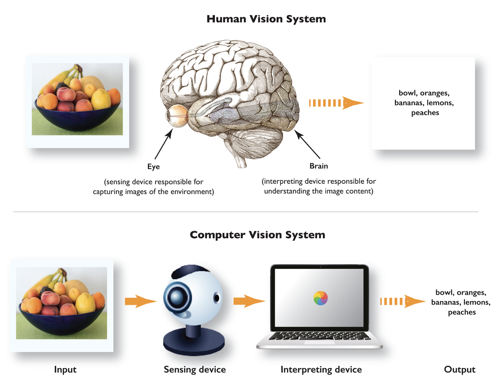
Computer vision in modern imaging is most effectively demonstrated through Computer-Aided Diagnosis (CAD) systems used in oncology screening. These systems leverage deep learning architectures, specifically Vision Transformers and Convolutional Neural Networks, to analyze high-resolution medical images like CT scans and MRIs, identifying malignant tissues with higher sensitivity and speed than traditional manual review.
The Convergence of Computer Vision and Modern Medical Imaging
By 2026, the field of modern imaging has moved beyond simple pixel capture to an integrated "intelligent sensor" paradigm. Computer vision (CV) is no longer an optional post-processing step; it is an intrinsic component of the imaging pipeline. In the context of oncology—specifically lung and breast cancer screening—CV provides the analytical layer that transforms raw volumetric data into actionable clinical insights.
Modern imaging relies on high-dimensional data. A single thoracic CT scan can generate hundreds of individual slices. For a human radiologist, the cognitive load required to maintain 100% accuracy across a full workday is immense. Computer vision serves as a "first reader" or a "concurrent reader," flagging suspicious regions of interest (ROIs) that may be smaller than two millimeters or obscured by dense physiological structures.
The primary problem solved by this CV example is the diagnostic bottleneck. By automating the detection of anomalies, CV allows specialists to focus on interpretation and treatment planning rather than the tedious task of searching for microscopic lesions.
Architectural Framework of Modern CV Systems in 2026
The technical sophistication of computer vision in 2026 is defined by the shift from purely Convolutional Neural Networks (CNNs) to Hybrid Vision Transformers (ViTs). While CNNs are excellent at capturing local patterns (like the edges of a tumor), Transformers excel at understanding global context—how a specific node relates to the rest of the organ system.
1. Data Acquisition and Normalization
Modern imaging sensors in 2026 utilize Adaptive Sensing, where the CV model provides real-time feedback to the imaging hardware. For example, if a model detects a potential anomaly during a low-dose CT scan, it can signal the hardware to increase the resolution in that specific spatial coordinate instantly. This reduces overall radiation exposure while maximizing detail where it matters most.
2. The Segmentation Pipeline
Once an image is captured, the CV system performs Semantic Segmentation. This involves labeling every pixel (or voxel in 3D) as a specific tissue type. In modern oncology, the U-Net++ architecture or its 2026 successors are used to delineate the boundaries of a tumor. Accurate segmentation is critical because it allows for the calculation of tumor volume, a key metric in assessing the efficacy of chemotherapy.
3. Feature Extraction and Classification
After segmentation, the system extracts high-level features. Unlike older "hand-crafted" features (where engineers told the computer to look for "roundness"), modern CV uses Representation Learning. The model learns to identify complex textures, vascular patterns, and density gradients that are invisible to the human eye. This is often referred to as Radiomics.
Step-by-Step Implementation of a CAD System
Implementing a computer vision system in a modern clinical environment involves a rigorous multi-stage process to ensure patient safety and data integrity.
Stage 1: Pre-processing and Denoising
Raw medical images are often "noisy" due to patient movement or hardware limitations. Modern systems use Generative Adversarial Networks (GANs) or Diffusion Models to denoise images without losing critical clinical detail. This step ensures that the downstream model is analyzing the truest possible representation of the anatomy.
Stage 2: Automated ROI Detection
The system scans the entire volume using a "sliding window" or an "anchor-based" approach. In 2026, Zero-Shot Learning models are frequently employed, allowing the system to flag "unusual" patterns even if it hasn't been explicitly trained on that specific rare subtype of cancer.
Stage 3: Quantitative Analysis
Once a lesion is detected, the CV system performs automated measurements. It calculates the RECIST (Response Evaluation Criteria in Solid Tumors) scores automatically. Traditionally, a radiologist would use digital calipers to measure a tumor manually; in 2026, the CV model provides a 3D volumetric measurement that is significantly more accurate for tracking disease progression.
Stage 4: Clinician Verification and Human-in-the-Loop
The final output is presented to the radiologist via an AI-augmented interface. The system highlights the ROI and provides a "confidence score" and an "explainability map" (often using SHAP or Integrated Gradients). This ensures the physician understands why the model flagged a specific area, maintaining the human-in-the-loop requirement for medical diagnostics.
Comparing Traditional vs. Modern CV Imaging (2026 Standards)
The transition from 2020-era technology to 2026-era modern imaging represents a leap in both hardware capabilities and algorithmic efficiency.
| Feature | Traditional CV (Circa 2020) | Modern CV (2026) |
|---|---|---|
| Model Architecture | Primarily 2D CNNs | 3D Vision Transformers (ViTs) |
| Detection Speed | Minutes per scan | Near real-time (Seconds) |
| Data Requirements | Large labeled datasets | Small-data/Self-supervised learning |
| Explainability | "Black Box" results | Heatmaps & textual reasoning |
| Hardware Integration | Post-capture analysis | Real-time sensor feedback |
| Diagnostic Accuracy | High false-positive rates | Precision-tuned low-latency inference |
| Regulatory Status | Emerging SaMD | Standard of Care (FDA/CE Integrated) |
Hardware and Infrastructure Requirements
To run a modern computer vision example in imaging, the infrastructure must handle massive datasets with low latency. By 2026, the hardware requirements have shifted toward Edge-Cloud Hybrids.
- Inference Hardware: Hospitals now utilize specialized AI accelerators like the NVIDIA Blackwell-series or specialized TPU (Tensor Processing Unit) clusters. These allow for the 3D processing of a 5GB CT study in under 10 seconds.
- Data Standards: Compliance with DICOM (Digital Imaging and Communications in Medicine) remains mandatory, but modern systems also integrate HL7 FHIR (Fast Healthcare Interoperability Resources) to pull patient history, which the CV model uses as metadata to inform its diagnostic confidence.
- Connectivity: 6G and high-speed fiber-optic intra-hospital networks are necessary to move high-fidelity volumetric data from the scanner to the inference engine without degradation.
Challenges, Troubleshooting, and Material Limits
Despite the advancements in 2026, computer vision in imaging is not without its limitations. Practitioners must be aware of several critical failure points.
Data Drift and Model Decay
A model trained on one manufacturer's MRI machine (e.g., Siemens) may perform poorly on a different machine (e.g., GE) due to subtle differences in sensor calibration. This is known as Domain Shift. Modern systems troubleshoot this through Continuous Monitoring, where the system's performance is audited monthly against biopsy results to ensure accuracy hasn't "drifted."
False Positives and "Over-Diagnosis"
One of the primary risks of high-sensitivity CV is the detection of benign lesions that would never have caused the patient harm. This leads to unnecessary biopsies and patient anxiety. In 2026, we address this by integrating Multi-modal AI, where the CV system analyzes the image alongside the patient's genetic markers and blood-work (Liquid Biopsies) to filter out clinically insignificant findings.
Legal and Ethical Guardrails
As of 2026, the legal framework for Software as a Medical Device (SaMD) is strictly enforced by agencies like the U.S. Food and Drug Administration (FDA). Developers must prove that their computer vision algorithms are free from "algorithmic bias"—ensuring the model performs equally well across different ethnicities, ages, and genders.
The Future of the "Example": From Detection to Prediction
The "example" of oncology screening is currently evolving from detection to prediction. In 2026, researchers are using CV to perform Predictive Phenotyping. By analyzing the visual texture of a tumor, computer vision can predict whether a specific immunotherapy drug will be effective before the treatment even begins. This moves modern imaging from a diagnostic tool to a cornerstone of Precision Medicine.
Furthermore, Federated Learning has become the standard for training these models. Instead of hospitals sharing sensitive patient images, the model travels to the data. It learns from a hospital’s local images and then shares only the mathematical "weights" with a central server. This preserves privacy while allowing the CV model to learn from millions of diverse cases globally.
Frequently Asked Questions
Can computer vision replace a radiologist in 2026?
No, computer vision cannot replace a radiologist. In 2026, CV is viewed as an "intelligence augment." While the AI is superior at pattern matching and finding tiny anomalies, the radiologist provides the necessary clinical context, ethical judgment, and patient communication that a machine cannot replicate. The goal is to automate the "search" so the human can focus on the "solution."
What is the difference between "Image Processing" and "Computer Vision" in modern imaging?
Image processing involves transforming an image to enhance it (e.g., adjusting contrast or sharpening edges). Computer vision goes a step further by interpreting the image content. If you use a filter to make a tumor clearer, that is image processing; if the computer tells you "there is a 92% chance this is a Stage II adenocarcinoma," that is computer vision.
How does "Explainable AI" (XAI) work in medical imaging?
XAI provides a rationale for a model's decision. In imaging, this often takes the form of a "Saliency Map," which highlights the specific pixels the model used to make its classification. If a model flags a lung nodule, XAI shows the radiologist that the model is looking at the spiculated (jagged) edges of the growth, which are a known indicator of malignancy.
Is computer vision in imaging secure from hackers?
Cybersecurity is a major focus in 2026. "Adversarial Attacks" are a known threat where hackers could theoretically introduce invisible noise into an image to trick the CV model into misdiagnosing a patient. To counter this, modern imaging pipelines use End-to-End Encryption and Adversarial Training, where the model is taught to recognize and ignore malicious digital noise.
What hardware is required for a hospital to implement these CV systems?
By 2026, most hospitals do not require on-site supercomputers. Instead, they use "AI-ready" imaging machines that connect to secure, localized medical clouds. However, they do need high-throughput networking (at least 10Gbps) and specialized diagnostic displays capable of rendering the high-resolution AI overlays accurately.
Practical Recommendation for Implementation
For healthcare institutions or developers looking to integrate computer vision into their imaging workflows in 2026, the primary focus should be on Interoperability. Ensure that your CV models are compliant with the latest HL7 FHIR standards and the DICOM-web protocol.
Start by deploying CV as a "shadow" system—allowing it to run in the background of your current workflow. Compare its findings with your existing manual reviews for a period of 6 to 12 months. This "Silent Trial" period is essential for building trust among clinical staff and identifying any domain-specific biases before the system is used to influence actual patient care decisions.