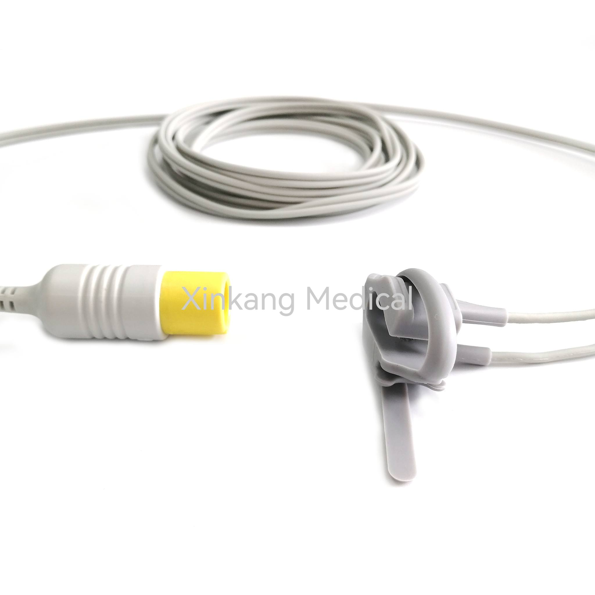
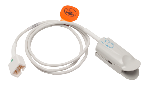
SpO2 stands for peripheral capillary oxygen saturation, an estimate of the amount of oxygen bound to hemoglobin in the blood relative to the total amount of hemoglobin that can bind, measured via a non-invasive sensor placed on the skin. This metric provides a rapid, painless indicator of how effectively the cardiovascular and respiratory systems are delivering oxygen to the peripheral tissues throughout the body.
Introduction to Pulse Oximetry and the SpO2 Acronym
In modern clinical practice and consumer health tracking, healthcare providers and patients frequently encounter medical acronyms that summarize complex physiological processes. Among the most ubiquitous of these terms is SpO2. Understanding this abbreviation requires examining both its individual linguistic components and the physiological mechanisms it evaluates.
The letter "S" typically stands for saturation, denoting the extent to which a specific molecule is occupied by another substance. The lowercase "p" stands for peripheral, which differentiates estimations taken through peripheral capillary beds—such as a fingertip, toe, or earlobe—from direct arterial blood gas (ABG) measurements taken via a needle puncture in an artery. The "O2" represents molecular oxygen. Together, the term describes the percentage of oxygen-saturated hemoglobin relative to total functional hemoglobin in peripheral blood vessels.
Breaking Down the Anatomy of the Term
To fully grasp the definition of the SpO2 medical abbreviation, one must look at how blood transports oxygen. Red blood cells contain hemoglobin, a specialized iron-containing protein that binds to oxygen molecules in the lungs and releases them as blood circulates through tissue beds.
- Hemoglobin Saturation: When all available binding sites on hemoglobin molecules are occupied by oxygen, the blood is fully saturated.
- Peripheral Measurement: Peripheral sensors rely on light absorption characteristics to estimate this saturation level without drawing blood.
- Fractional vs. Functional: While laboratory co-oximeters measure fractional saturation (including dysfunctional hemoglobins like carboxyhemoglobin and methemoglobin), standard pulse oximetry focuses primarily on functional saturation—the ratio of oxyhemoglobin to deoxyhemoglobin.
The Physiological Basis of Blood Oxygen Saturation
Oxygen is essential for cellular respiration and energy production in humans. Ambient air enters the lungs during inhalation, passes across the alveolar-capillary membrane, and dissolves into the bloodstream. Here, the vast majority of oxygen binds to hemoglobin molecules within erythrocytes, while a small fraction remains dissolved directly in the plasma.
When clinicians reference SpO2, they are quantifying the efficiency of this gas exchange process. A normal, healthy individual breathing ambient air at sea level typically exhibits an SpO2 reading between 95% and 100%. Values consistently falling below this baseline may indicate underlying pulmonary or cardiovascular dysfunction, prompting further diagnostic evaluation.
Factors Influencing SpO2 Readings
Multiple physiological and environmental variables can influence the accuracy and interpretation of an SpO2 reading. Understanding these factors prevents misinterpretation by patients and medical professionals alike:
- Perfusion Status: Because pulse oximeters rely on pulsatile blood flow to detect arterial hemoglobin, conditions causing poor peripheral circulation—such as severe hypothermia, vasoconstriction, or low cardiac output—can diminish signal quality.
- Skin Pigmentation and Thickness: Variations in melanin concentration, thick skin, or calluses at the sensor site can alter light absorption, though modern devices utilize advanced signal processing algorithms to minimize these discrepancies.
- Hemoglobin Variants: The presence of abnormal hemoglobin types, such as carboxyhemoglobin (resulting from carbon monoxide poisoning) or methemoglobin, can cause pulse oximeters to display misleadingly high readings because these variants absorb light similarly to oxyhemoglobin.
- Motion Artifact: Physical movement of the patient's finger or limb during measurement can introduce noise into the optical signal, leading to erroneous or fluctuating values.
How Pulse Oximetry Works Technologically
The acquisition of an SpO2 measurement relies on a technology known as pulse oximetry, which uses spectrophotometry and photoplethysmography. A typical pulse oximeter probe features two or more light-emitting diodes (LEDs) and a photodetector positioned on opposite sides of a tissue bed, most commonly a fingertip.
The device emits light at specific wavelengths: red light (typically around 660 nanometers) and infrared light (typically around 940 nanometers). Oxyhemoglobin and deoxyhemoglobin absorb these two wavelengths of light differently. Oxyhemoglobin absorbs more infrared light and allows more red light to pass through, whereas deoxyhemoglobin absorbs more red light and allows more infrared light to pass through.
By measuring the differential absorption of these light beams during the pulsatile surge of arterial blood, the device calculates the ratio of oxyhemoglobin to total hemoglobin. This calculation is processed by an internal microchip and displayed on a digital screen as a percentage, accompanied by a pulse rate and a plethysmographic waveform.
Clinical Interpretation of SpO2 Values
Interpreting SpO2 values requires context, including the patient's baseline health status, age, underlying chronic conditions, and environmental factors such as altitude. Clinicians use specific thresholds to guide medical decision-making.
| SpO2 Range (%) | Clinical Interpretation | Potential Actions & Considerations |
|---|---|---|
| 95% – 100% | Normal oxygen saturation for healthy individuals. | Standard baseline; no intervention required unless acute symptoms are present. |
| 90% – 94% | Mild hypoxemia or normal baseline for chronic conditions. | Monitor closely; acceptable for stable patients with chronic obstructive pulmonary disease (COPD) depending on physician targets. |
| 85% – 89% | Moderate hypoxemia requiring clinical attention. | Requires medical evaluation; supplemental oxygen may be indicated based on arterial blood gas correlation. |
| Below 85% | Severe hypoxemia representing a medical emergency. | Immediate intervention required, including high-flow supplemental oxygen and emergency medical services. |
Note: Individual targets vary. Patients with chronic respiratory diseases often maintain lower baseline SpO2 ranges as established by their treating pulmonologist.
SpO2 Versus SaO2: Understanding the Distinction
A common point of confusion among students and healthcare consumers is the difference between SpO2 and SaO2. While both terms describe arterial oxygen saturation, their methods of acquisition and exact definitions differ significantly.
- SaO2 (Arterial Oxygen Saturation): This is measured directly by analyzing an arterial blood gas (ABG) sample drawn from an artery via a syringe and needle. It represents the precise percentage of hemoglobin binding sites occupied by oxygen in an actual blood specimen.
- SpO2 (Peripheral Capillary Oxygen Saturation): This is estimated non-invasively via a pulse oximeter placed on the skin. It relies on optical sensors rather than chemical analysis of blood chemistry.
While SpO2 serves as a reliable, rapid proxy for SaO2 in most clinical scenarios, healthcare providers may order an ABG (yielding SaO2, PaO2, pH, and carbon dioxide levels) when exact acid-base balance and dissolved gas pressures are required.
Applications in Clinical Settings and Home Health
The utility of tracking SpO2 spans diverse medical environments, from critical care units to home monitoring programs.
Acute Care and Surgery
In operating rooms and emergency departments, continuous pulse oximetry is a standard monitoring modality. Anesthesiologists and nurses rely on real-time SpO2 trends to detect early signs of hypoxia during sedation, airway manipulation, or critical illness before clinical cyanosis (bluish discoloration of the skin) becomes visible.
Chronic Disease Management
Patients living with chronic respiratory conditions, such as chronic obstructive pulmonary disease (COPD), pulmonary fibrosis, or severe asthma, frequently use home pulse oximeters. These devices help patients track their daily status, evaluate the effectiveness of prescribed oxygen therapy, and recognize acute exacerbations early.
High-Altitude Travel and Athletics
Mountaineers, pilots, and elite athletes monitor SpO2 to assess physiological adaptation to hypobaric hypoxia at high altitudes. Acclimatization involves compensatory mechanisms like increased ventilation and red blood cell production, which can be tracked over time using portable oximeters.
Limitations and Potential Pitfalls of Pulse Oximeters
Despite their widespread utility, consumer and clinical pulse oximeters have inherent limitations that users must understand to avoid diagnostic errors.
- Lag Time: Pulse oximetry reflects changes in oxygen saturation rapidly, but severe hypoventilation or apnea may alter carbon dioxide levels in the blood before oxygen saturation drops significantly.
- Nail Polish and Artificial Nails: Dark nail polishes (such as black, blue, or dark green) and acrylic nails can absorb or block the light wavelengths emitted by the sensor, leading to erroneously low readings.
- Ambient Light Interference: Bright operating room lights, sunlight, or phototherapy lamps can overwhelm the photodetector if the sensor is not properly shielded.
- Motion and Low Perfusion Artifacts: Shivering, tremors, or poor peripheral blood flow can cause the device to display erratic numbers or fail to register a pulse entirely.
Frequently Asked Questions About SpO2
What is the difference between SpO2 and pulse rate?
SpO2 measures the percentage of oxygen-saturated hemoglobin in the blood, whereas pulse rate measures how many times the heart beats per minute. Most pulse oximeters display both metrics simultaneously, but they evaluate entirely different physiological parameters.
Can an ordinary smartwatch accurately measure SpO2?
Many consumer smartwatches and fitness trackers feature optical sensors capable of estimating SpO2. While these devices are useful for general wellness tracking and trend analysis, they are not FDA-cleared diagnostic medical devices and should not be used to self-diagnose or manage medical conditions without professional oversight.
What should I do if my SpO2 reading drops below 95%?
If you observe a low reading, first check for common interference factors: warm your hands if they are cold, remove any dark nail polish, and remain still while taking a new measurement. If low readings persist—particularly below 92% or when accompanied by shortness of breath, chest pain, or confusion—seek immediate medical evaluation.
Does cold weather affect SpO2 readings?
Yes. Exposure to cold temperatures causes peripheral vasoconstriction, reducing blood flow to the fingers and toes. This lack of adequate pulsatile blood flow can make it difficult for a pulse oximeter to obtain an accurate reading, often resulting in artificially low values or error codes.
Why do readings fluctuate from second to second?
Pulse oximeters calculate saturation based on pulsatile blood flow with each heartbeat. Minor physiological variations in blood volume, breathing depth, and slight finger movements can cause the digital display to fluctuate by one to two percentage points continuously.
Conclusion and Next Actions
The SpO2 medical abbreviation stands for peripheral capillary oxygen saturation, serving as an indispensable, non-invasive metric for assessing blood oxygenation levels. Whether utilized in high-acuity hospital settings or monitored at home for chronic respiratory management, understanding what this percentage represents empowers patients and clinicians to recognize normal physiology versus acute distress. If you are monitoring your SpO2 at home for a medical condition, maintain a symptom log, discuss target ranges with your physician, and consult qualified healthcare professionals for any persistent or concerning readings.