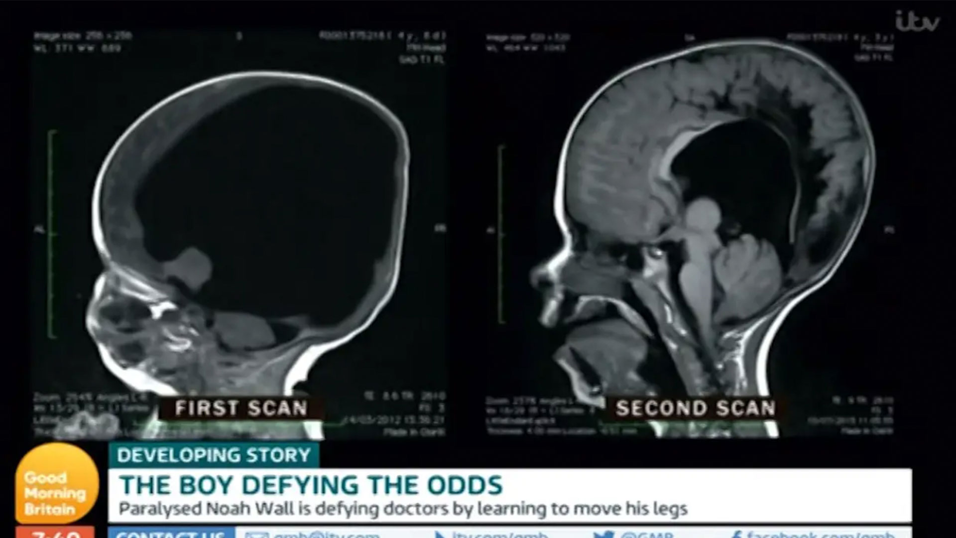
A person cannot survive entirely without a brain, as the brainstem is required to regulate autonomic functions like breathing and heart rate. However, individuals can live with significant portions of their brain missing due to conditions such as hydranencephaly or extreme hydrocephalus. In these rare cases, the brainstem remains functional while the cerebral hemispheres are largely absent or displaced by fluid.
The concept of a person "without a brain" often surfaces in medical literature not as a total biological void, but as a spectrum of severe neurological conditions. While popular media occasionally highlights sensational stories of individuals functioning with "no brain," the clinical reality is far more complex. These cases challenge our traditional understanding of neuroanatomy, consciousness, and the limits of human biology. As of 2026, advances in neuroimaging and neonatal care have allowed us to better understand how the human body adapts when the most vital organ is severely compromised or partially absent from birth.
Clinical Definitions: Anencephaly vs. Hydranencephaly
To discuss a person without a brain accurately, we must first distinguish between the various medical conditions that involve the absence of brain tissue. These conditions occur during embryonic development and are generally classified under the umbrella of neural tube defects or vascular disruptions.
Anencephaly: The Absence of the Major Brain
Anencephaly is one of the most severe forms of neural tube defects. It occurs when the upper part of the neural tube fails to close, resulting in the absence of a major portion of the brain, skull, and scalp. In most cases, the forebrain (the cerebrum) and the cerebellum are missing.
Individuals born with anencephaly are usually blind, deaf, and unable to feel pain. While the brainstem may be partially present, providing enough reflex action to allow for breathing or response to touch, the lack of a functioning cerebrum means that higher-order consciousness is impossible. According to the Centers for Disease Control and Prevention (CDC), most infants born with anencephaly do not survive more than a few hours or days after birth.
Hydranencephaly: Fluid in Place of the Hemispheres
Hydranencephaly is often what people refer to when they speak of someone "living without a brain." Unlike anencephaly, where the skull is missing, a child with hydranencephaly is born with a seemingly normal head shape. However, inside the skull, the cerebral hemispheres are almost entirely absent, replaced by sacs of cerebrospinal fluid (CSF).
The cause is believed to be a massive vascular insult, such as a stroke or blockage of the carotid arteries, occurring in utero after the basic structure of the brain has started to form. Because the brainstem and cerebellum are usually spared, these children can breathe, swallow, and maintain a sleep-wake cycle. Some children with hydranencephaly have lived into their teens or even early twenties, though they face profound developmental delays and physical disabilities.
The Case of the "Missing" Brain: Extreme Hydrocephalus
One of the most famous cases in neurology involves a 44-year-old French civil servant who went to the doctor in 2007 complaining of mild leg weakness. When doctors performed an MRI, they were shocked to find that his brain was almost entirely missing. His cranial cavity was filled with fluid, with only a thin perimeter of actual brain tissue lining the skull.
How Function Persists with Minimal Tissue
This man’s condition was the result of long-term, untreated hydrocephalus (water on the brain). As fluid built up over decades, it slowly compressed his brain matter against the skull. Despite having roughly 90% of his brain displaced by fluid, the man was a married father of two and worked as a civil servant. His IQ was measured at 75—below the average of 100 but significantly higher than what one would expect given his scans.
This case, published in The Lancet, remains a cornerstone of discussions regarding neuroplasticity. It suggests that if the brain is compressed slowly enough over time, the remaining tissue can sometimes take over the functions of the lost areas. The brain’s ability to "rewire" itself allows for functional survival even when the physical mass of the organ is severely diminished.
The Essential Role of the Brainstem
The reason a person can survive with missing cerebral hemispheres but not a "complete" absence of the brain lies in the brainstem. Located at the base of the brain and connecting to the spinal cord, the brainstem is the command center for the body’s most basic life-support systems.
- The Medulla Oblongata: Controls autonomic functions such as breathing, heart rate, and blood pressure.
- The Pons: Acts as a bridge between various parts of the brain and plays a role in sleep and respiratory rhythm.
- The Midbrain: Involved in motor movement and auditory/visual processing.
As long as the brainstem is intact and functioning, the body can technically remain alive without the "higher" functions of the cerebrum, such as thought, language, and voluntary movement. This state is often referred to as a vegetative state or, in the case of congenital absence, "permanent unconsciousness."
Neuroplasticity: The Brain’s Ability to Adapt
The persistence of function in individuals with severe brain loss is attributed to neuroplasticity. This is the nervous system’s ability to change its activity in response to intrinsic or extrinsic stimuli by reorganizing its structure, functions, or connections.
In cases of extreme hydrocephalus or hemispherectomies (the surgical removal of half the brain), the remaining neurons undergo a process of massive reorganization. This includes:
- Synaptogenesis: Creating new connections between existing neurons.
- Functional Reassignment: One part of the brain learning to perform tasks originally assigned to a missing part.
- Axonal Sprouting: Undamaged axons growing new nerve endings to reconnect neurons whose links were severed.
While neuroplasticity has limits—it cannot create complex thought where no cortex exists—it explains why some individuals with significant brain damage or missing segments can still walk, talk, or perform basic tasks.
Comparing Severe Brain Anomalies
The following table summarizes the differences between the conditions most often associated with the concept of a person living "without a brain."
| Condition | Primary Brain Absence | Skull/Appearance | Life Expectancy | Functional Ability |
|---|---|---|---|---|
| Anencephaly | Major parts of brain and skull missing | Visible opening/absence | Hours to days | Minimal reflexes only |
| Hydranencephaly | Cerebral hemispheres replaced by fluid | Appears normal or slightly enlarged | Days to years | Basic reflexes; no higher cognition |
| Extreme Hydrocephalus | Brain tissue compressed to a thin layer | May be enlarged (macrocephaly) | Can be normal with treatment | Variable; can include near-normal IQ |
| Brain Death | Total cessation of all brain/brainstem function | Normal | Minutes without life support | None; legal definition of death |
Ethical and Legal Definitions of Death
The discussion of living without a brain inevitably leads to the legal definition of death. In most jurisdictions, including the United States under the Uniform Determination of Death Act (UDDA), death is defined as either the irreversible cessation of circulatory and respiratory functions or the irreversible cessation of all functions of the entire brain, including the brainstem.
A person whose brainstem still functions is legally alive, even if they lack a cerebrum and the capacity for consciousness. This creates significant ethical challenges in neonatal care and end-of-life decisions. For instance, should an infant with anencephaly be considered an organ donor while their heart is still beating? These questions continue to be debated among medical ethicists and legal experts in 2026 as medical technology makes it possible to sustain the body longer than ever before.
Diagnostic Tools and Modern Detection
In 2026, the detection of these conditions has become highly sophisticated. Prenatal screening is the primary method for identifying severe brain anomalies before birth.
- Ultrasound: High-resolution 3D and 4D ultrasounds can detect anencephaly as early as the first trimester.
- Fetal MRI: When an ultrasound is inconclusive, a fetal MRI provides detailed images of the developing brain's structure, allowing doctors to distinguish between hydrocephalus and hydranencephaly.
- Alpha-Fetoprotein (AFP) Screening: High levels of AFP in the mother's blood often indicate a neural tube defect like anencephaly.
Early diagnosis is crucial for family planning and for determining the level of palliative care required at birth. For adults, MRI and CT scans remain the gold standard for identifying hidden anomalies, as seen in the case of the French civil servant.
Frequently Asked Questions
Can a person feel pain without a brain?
If the person has a functioning brainstem but lacks a cerebral cortex (as in hydranencephaly), they may exhibit "pain behaviors" such as crying or withdrawing from a stimulus. However, most neuroscientists believe that the experience of pain—the conscious awareness of it—requires the somatosensory cortex. Without a cortex, these responses are likely subcortical reflexes rather than conscious suffering.
How long can someone with hydranencephaly live?
While many infants with hydranencephaly die within their first year, there are documented cases of individuals living into their 20s. Survival depends on the stability of their autonomic functions and the prevention of secondary complications like pneumonia or severe seizures.
Is "brain death" the same as having no brain?
No. Brain death is a clinical diagnosis meaning the entire brain, including the brainstem, has permanently stopped functioning. A person with anencephaly or hydranencephaly has a functioning brainstem and is technically alive, whereas a brain-dead person is legally and biologically dead, even if a ventilator keeps their heart beating.
Can a person with half a brain live normally?
Yes. This is often seen in patients who undergo a hemispherectomy to treat severe epilepsy. If the surgery is performed at a young age, the remaining half of the brain can often adapt to handle the functions of both sides, allowing the person to lead a relatively normal life with only minor physical limitations.
Are there any treatments for these conditions?
Anencephaly and hydranencephaly have no known cure. Treatment is palliative, focusing on comfort and managing symptoms. In cases of hydrocephalus, the insertion of a shunt to drain excess fluid can prevent the destruction of brain tissue and allow for normal or near-normal development.
Practical Next Actions for Affected Families
If a family receives a diagnosis of a severe brain anomaly during pregnancy, the first step is to consult with a team of specialists, including a maternal-fetal medicine specialist, a pediatric neurosurgeon, and a genetic counselor.
- Seek a Second Opinion: Because these conditions are rare, it is vital to have imaging reviewed by specialists at a major neurological center.
- Palliative Care Planning: For conditions like anencephaly, developing a perinatal hospice or palliative care plan can help families focus on the short time they will have with their child.
- Support Networks: Organizations such as the National Institute of Neurological Disorders and Stroke (NINDS) provide resources and connections to support groups for families dealing with rare neurological disorders.
While the prospect of a person living "without a brain" sounds like science fiction, it is a poignant and complex medical reality. Whether through the tragedy of congenital defects or the marvel of neuroplasticity, these cases remind us that the human body is remarkably resilient and that our understanding of the "seat of the soul" is still evolving.