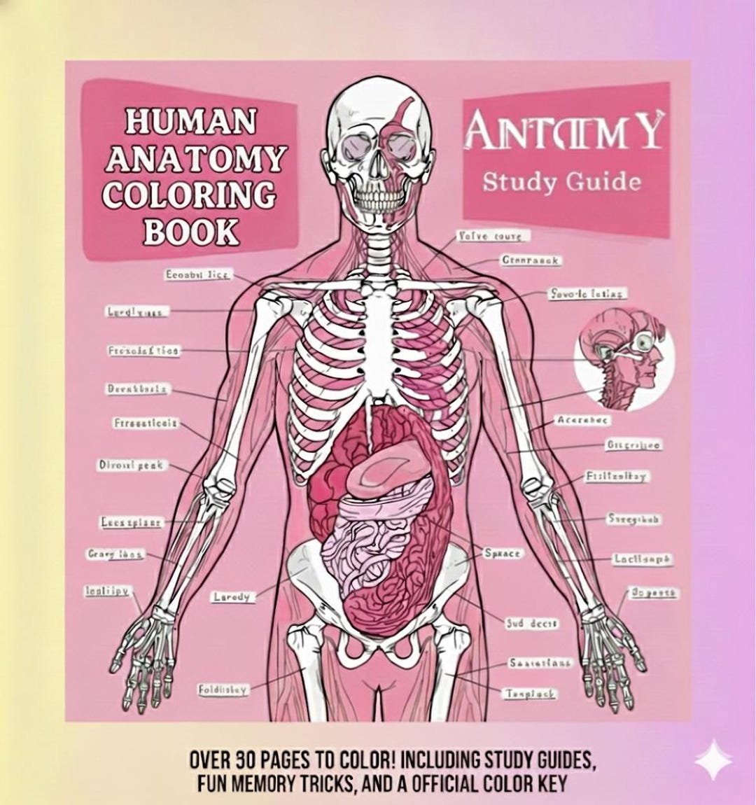
Anatomy coloring book PDF resources provide downloadable, printable, or tablet-compatible line illustrations designed to help medical, nursing, and biology students learn human anatomy through active recall and visual engagement. Sourcing legitimate files involves accessing open educational resources (OER), museum archives, and authorized publisher digital platforms to obtain clear, high-resolution diagrams.
The Cognitive Science Behind Anatomy Coloring
Studying complex anatomical structures requires moving beyond passive reading or static diagram review. Human anatomy demands an understanding of spatial relationships, anatomical orientation (such as anterior, posterior, medial, and lateral aspects), and functional systems. Coloring anatomical diagrams engages multiple sensory modalities, creating stronger memory traces than passive viewing alone.
Educational psychologists refer to this effect through dual-coding theory, originally proposed by Allan Paivio. Dual-coding theory demonstrates that the brain processes information through visual and verbal channels simultaneously. When a student reads an anatomical term (e.g., brachiocephalic artery), locates the corresponding vessel on an uncolored line drawing, and manually fills that structure with a distinct color, the brain links textual, spatial, and motor pathways.
- Visual Channel (Diagram & Color)
- Verbal Channel (Term & Definition)
- Combined Mental Model & Active Recall
Furthermore, anatomy coloring acts as a form of active recall. Unlike pre-colored textbook images where boundaries and labels are already highlighted, an uncolored PDF workbook forces the learner to identify structural borders, distinguish origin and insertion points of muscles, and trace vascular or neural pathways independently. This process builds micro-decisions into study sessions:
- Is this structure superficial or deep to the adjacent muscle?
- Does this nerve pass anterior or posterior to the medial epicondyle?
- Which organ system does this specific capillary bed serve?
By resolving these spatial queries during the act of coloring, students reduce cognitive overload during high-stakes practical exams, pin tests, and clinical rotations.
Where to Find Legitimate Anatomy Coloring Book PDFs
Finding reliable, high-resolution anatomy coloring book PDFs requires navigating between free open-access archives, institutional open educational resources (OER), and official publisher digital downloads. Because copyright restrictions protect proprietary commercial workbooks, students should prioritize verified legal sources that offer clear line art suitable for printing or digital annotation.
- Anatomy PDF Sources
- Open Educational Resources (OER) & University Repositories
- Institutional Archives (NYAM "Color Our Collections")
- Publisher Digital Companions (Elsevier Evolve, Pearson)
- Open-Access Biological Libraries (Biodiversity Heritage Library)
Open Educational Resources (OER) and University Repositories
Many universities and academic consortia publish open-access anatomy workbooks licensed under Creative Commons. These resources are specifically created for health science, kinesiology, and introductory biology courses.
- OpenStax Ancillary Materials: Rice University’s OpenStax provides free, open-source Anatomy and Physiology textbooks. Alongside the main texts, faculty and community contributors frequently share downloadable PDF line-art figures and workbook packets optimized for coloring and labeling.
- Institutional Digital Commons: University libraries—such as those at the University of Michigan, the University of Iowa, and the University of Utah—host open repository files created by faculty. These often include regional anatomy worksheets, neuroanatomy line diagrams, and histology tracing sheets formatted directly as PDF downloads.
- LibreTexts Medicine: The LibreTexts platform hosts collaborative, peer-reviewed medical and anatomical modules that feature printable black-and-white anatomical diagrams designed for active student annotation.
Historical and Medical Museum Archives
For historical, microscopic, and macro-anatomical line drawings, institutional libraries frequently digitize public-domain anatomical plates.
- New York Academy of Medicine (NYAM) – Color Our Collections: Every year, NYAM coordinates an international campaign where libraries, archives, and medical museums release free, downloadable PDF coloring books based on their rare collections. Participating institutions—such as the National Library of Medicine (NLM), the Royal College of Physicians, and the Francis A. Countway Library of Medicine at Harvard—provide detailed historical anatomical sketches, ranging from Vesalian structural plates to 19th-century neuroanatomical line art.
- Biodiversity Heritage Library (BHL): BHL offers millions of high-resolution, public-domain natural history and comparative anatomy illustrations. Users can export specific anatomical plates as high-grade PDFs suitable for physical printing.
Official Publisher Digital Workbooks and Student Companions
While complete commercial bestsellers—such as The Anatomy Coloring Book by Wynn Kapit and Lawrence M. Elson, or Netter's Anatomy Coloring Book by John T. Hansen—are protected by commercial copyright, publishers offer legal digital access points:
- Publisher Student Companions: Purchasing a physical or digital copy of authorized anatomy coloring books often includes an access code to publisher portals (such as Elsevier Evolve or Pearson Higher Ed). These portals frequently grant purchasers access to downloadable, full-sized PDF worksheets of every plate in the book, allowing students to print fresh practice sheets without damaging their physical copies.
- Digital eBook Platforms: Authorized digital textbook providers like VitalSource, RedShelf, and Inkling offer interactive PDF-based or ePub formats of standard coloring workbooks. These platforms allow direct digital drawing using styluses on tablets.
Comparative Evaluation of Anatomy PDF Sources
Selecting the right PDF resource depends on your academic focus, budget, and intended output method (physical printing vs. tablet annotation). The table below compares primary sources of anatomy coloring PDF files based on access, cost, line art style, and target audience.
| Resource Category | Primary Examples | Access & License | Line Art Format | Primary Audience & Best Use Case |
|---|---|---|---|---|
| Academic OER Workbooks | OpenStax A&P Worksheets, LibreTexts | Free (Creative Commons) | Vector & Clean Raster PDF | Undergraduate A&P, Nursing, Allied Health basic preparation |
| Publisher Digital Companions | Elsevier Evolve (Netter's), Pearson (Kapit & Elson) | Paid (Requires Book/Code Purchase) | High-DPI Digital PDF | Medical (MD/DO), Dental, Physical Therapy students needing clinical depth |
| Library & Museum Archives | NYAM "Color Our Collections", NLM Archives | Free (Public Domain / Open Access) | Historical Fine-Line Raster PDF | Advanced histology, neuroanatomy interest, historical anatomy study |
| Open University Repositories | University Repository Commons, Open Michigan | Free (Institutional License) | Clean Black-and-White Line PDFs | Targeted organ-system review, muscular/skeletal focus |
| Digital Tablet Worksheets | Specialized Etsy/Gumroad Medical Creators | Paid (Direct Digital Purchase) | Optimized PDF with Layers / Vector | iPad/Tablet users looking for pre-formatted digital notebook templates |
Technical Criteria for High-Quality Anatomy PDF Resources
Not all downloadable PDFs are created equal. Poorly scanned documents or compressed image files can create pixelated lines, muddy shading, and unreadable text labels that disrupt study. When evaluating an anatomy coloring book PDF, consider the following technical specifications.
Vector Graphics vs. High-DPI Raster Graphics
Anatomical line art generally comes in two digital image types:
- Vector PDF (Scalable Vector Graphics embedded in PDF): Vector files use mathematical formulas to draw lines. They can be zoomed in infinitely without losing crispness. These are ideal for studying micro-structures (such as the nephron of the kidney or the inner ear ossicles) on high-resolution displays or when printing enlarged poster sheets.
- Raster PDF (Bitmap Line Art): Raster PDFs consist of pixel grids. If sourcing raster PDFs, ensure the document has a minimum resolution of 300 DPI (dots per inch) at full page size (8.5" x 11" or A4). Scans below 150 DPI produce jagged, blurry lines when printed, making fine structures hard to delineate.
- Vector PDF (Mathematical paths): Infinite resolution, crisp lines at any zoom level
- Raster PDF (Pixel grid at 300+ DPI): High detail, fixed printable size without pixelation
- Low-Res Scan (< 150 DPI): Pixelated edges, illegible labels (Avoid for study)
Labeling Systems and Test Modes
Effective study PDFs usually incorporate structured labeling designs:
- Fully Labeled Master Sheets: Useful for initial study, orientation, and reference.
- Unlabeled/Blind Test Sheets: Feature lead lines pointing to structures with blank numbered keys, allowing students to test their active recall before checking the key.
- Layered PDFs: Advanced PDF documents allow users to toggle visibility layers on and off in software like Adobe Acrobat Reader, hiding or showing labels, lead lines, and background grids dynamically.
How to Integrate Anatomy PDFs into Medical Study Workflows
To maximize retention when using anatomy coloring PDFs, adopt a structured, systematic study routine rather than treating coloring as passive artwork.
1. Establish Standardized Color Schemes
Using consistent colors across different organ systems builds subconscious visual associations. For instance, medical education standardized conventions include:
- Red: Arteries, oxygenated blood flow, muscle bellies (or muscle origins).
- Blue: Veins, deoxygenated blood flow, cartilage, muscle insertions.
- Yellow: Peripheral nerves, cranial nerves, nerve tracts, adipose tissue.
- Green: Lymphatic vessels, lymph nodes, bile ducts.
- Purple/Brown: Digestive tract segments, glandular tissue, cross-sectional parenchyma.
Maintaining these color conventions across all your PDF worksheets helps your brain instantly categorize structures during timed examination scenarios.
2. Tablet Annotation Setup (iPad & Android)
If studying digitally on tablets using applications such as GoodNotes, Notability, Samsung Notes, or Procreate:
Import the PDF: Open the vector or high-DPI PDF directly into your note-taking application.
Use Pencil or Highlighter Tools set to Multiplier/Pass-Through Mode: Configure your digital stylus highlighter to an opacity level between 30% and 50%. This ensures that the digital ink lays behind the black vector line art rather than obscuring essential structural outlines.
Organize by System or Region: Create digital notebooks organized regionally (e.g., Head and Neck, Thorax, Upper Limb) or systemically (e.g., Musculoskeletal, Cardiovascular, Nervous System).
Import Anatomy PDF
Set Pencil/Highlighter Opacity (30-50%)
Apply Color Coding
Hide/Test Labels
3. Physical Printing and Paper Selection
If you prefer physical paper for kinesthetic learning:
- Paper Weight: Standard 20 lb printer paper easily bleeds through when using alcohol markers, gel pens, or heavy highlighters. Print your anatomy PDFs on 24 lb to 32 lb bright white paper or fine-tooth cardstock to prevent ink bleed and tearing.
- Binding: Use a three-ring binder or disc-bound notebook to keep sheets organized by region, allowing you to insert fresh, unlabeled test sheets opposite your fully colored master reference sheets.
Copyright, Licensing, and Avoiding Malware
Searching for free PDF downloads of commercial textbooks often leads to piracy sites, torrent repositories, or unverified file-sharing portals. Unauthorized uploads of copyrighted books carry significant drawbacks:
- Low Quality: Pirated PDFs are routinely compiled from low-quality, skewed flatbed scans with cropped margins, missing pages, or unusable low resolutions.
- Security Risks: Downloadable files from unverified file-sharing networks frequently embed malicious scripts, adware, or ransomware disguised as executable PDF extensions (
.pdf.exe). - Legal Issues: Distributing or downloading copyrighted material violates international intellectual property regulations and university academic honor codes.
Best Practice: Always verify that the host platform is an accredited educational institution, an established library archive, an open-access repository, or an authorized publisher portal. Look for clear licensing statements such as Creative Commons (CC BY-NC-SA) or Public Domain Mark 1.0.
Frequently Asked Questions
Are free anatomy coloring book PDFs legally available online?
Yes. Legitimate, free anatomy coloring PDFs are available through Open Educational Resources (OER) like OpenStax, university digital repositories, and historical archives participating in initiatives like the New York Academy of Medicine's "Color Our Collections." Commercial textbooks (e.g., Netter's or Kapit & Elson) require paid purchase or authorized publisher code access.
What paper type is best for printing anatomy coloring sheets?
Use 24 lb to 32 lb bright white printer paper or lightweight cardstock. Standard 20 lb copy paper easily bleeds when colored with markers, highlighters, or liquid ink pens, obscuring fine line art on the reverse side.
How do I color an anatomy PDF on an iPad without hiding the black lines?
In note-taking apps like GoodNotes or Notability, use the highlighter tool, which automatically sits behind printed lines. In art applications like Procreate or Adobe Photoshop, place your color layer below the imported line art layer, or set the line art layer's blend mode to Multiply.
Can anatomy coloring books replace traditional medical flashcards?
No, coloring books work best when paired alongside active recall tools like flashcards (e.g., Anki). Coloring builds initial spatial orientation and visual-motor recognition, while spaced-repetition flashcards reinforce long-term anatomical term retention and clinical facts.
How do I check if a PDF line drawing is high enough resolution to print clearly?
Open the PDF document on a computer screen and zoom in to 200% or 300%. If the line art remains sharp and crisp without pixelation or jagged edges, it is either a vector graphic or a high-DPI scan that will print clearly.
Practical Next Actions for Students
To build an efficient anatomy study workspace using PDF coloring resources:
- Source Verified Files: Download open-access foundational anatomical worksheets from OpenStax, LibreTexts, or your university library system. If using a major commercial text, redeem your publisher code to access printable digital supplements.
- Prepare Your Media: If using a digital tablet, set up color palettes matching standard medical conventions (red for arteries, blue for veins, yellow for nerves). If printing, stock your printer with 24 lb or 32 lb paper and gather dual-tip markers or colored pencils.
- Establish a Study Sequence: Read the corresponding anatomical chapter first, color the PDF worksheet while identifying key landmarks, and immediately create Anki cards for unlabeled diagrams to test your retention over time.