An astrocytoma is a type of brain tumor that originates in star-shaped glial cells called astrocytes, which support and protect neurons in the central nervous system. These tumors vary significantly in growth rate and severity, ranging from slow-growing benign lesions to highly aggressive, invasive malignancies that require urgent multimodal intervention.
What is an Astrocytoma?
Astrocytomas belong to a broader category of brain tumors known as gliomas. To understand an astrocytoma, one must first understand the role of the astrocyte. In a healthy brain and spinal cord, astrocytes perform critical "housekeeping" functions: they maintain the blood-brain barrier, provide nutrients to nervous tissue, and regulate the chemical environment necessary for synaptic transmission.
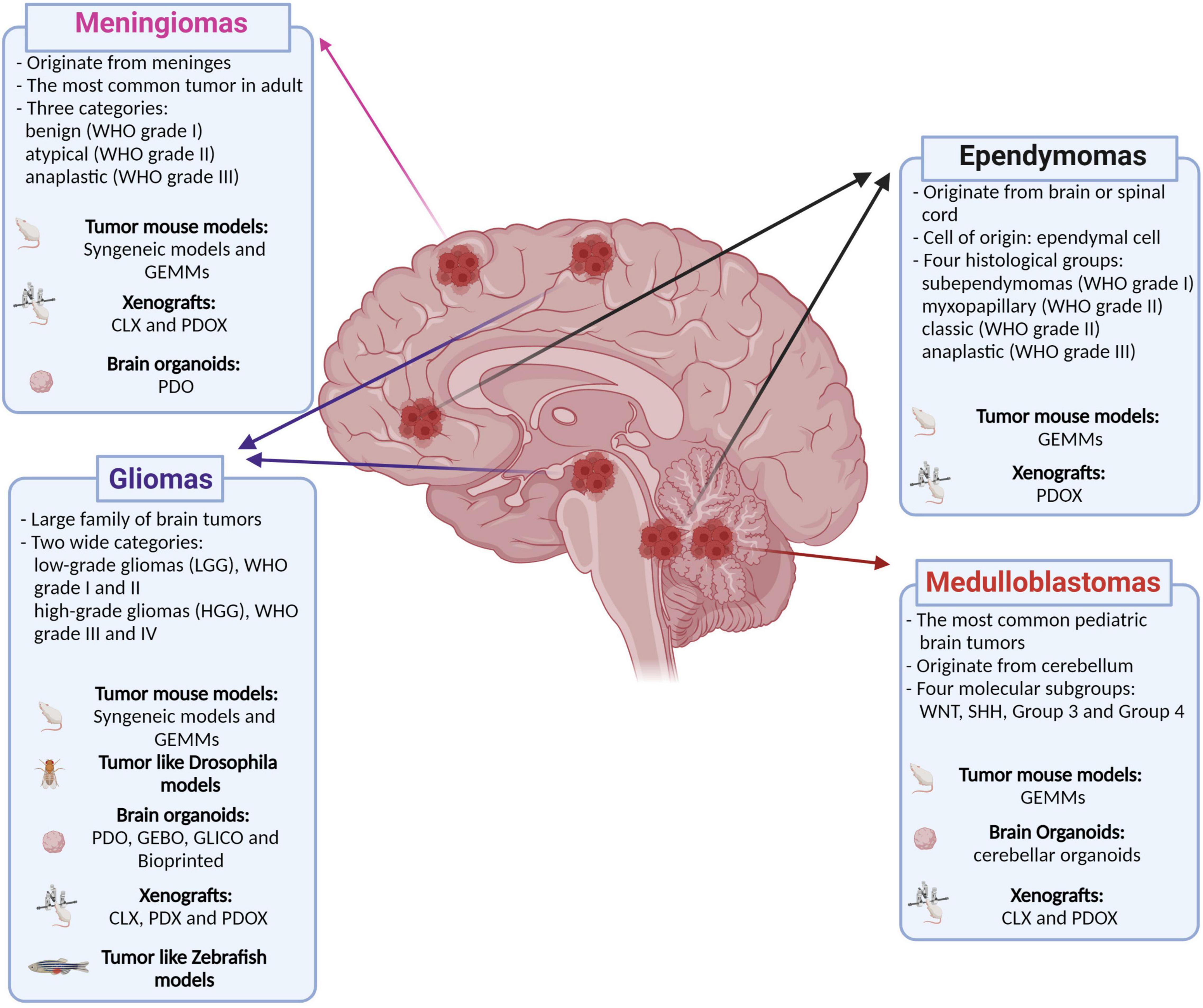
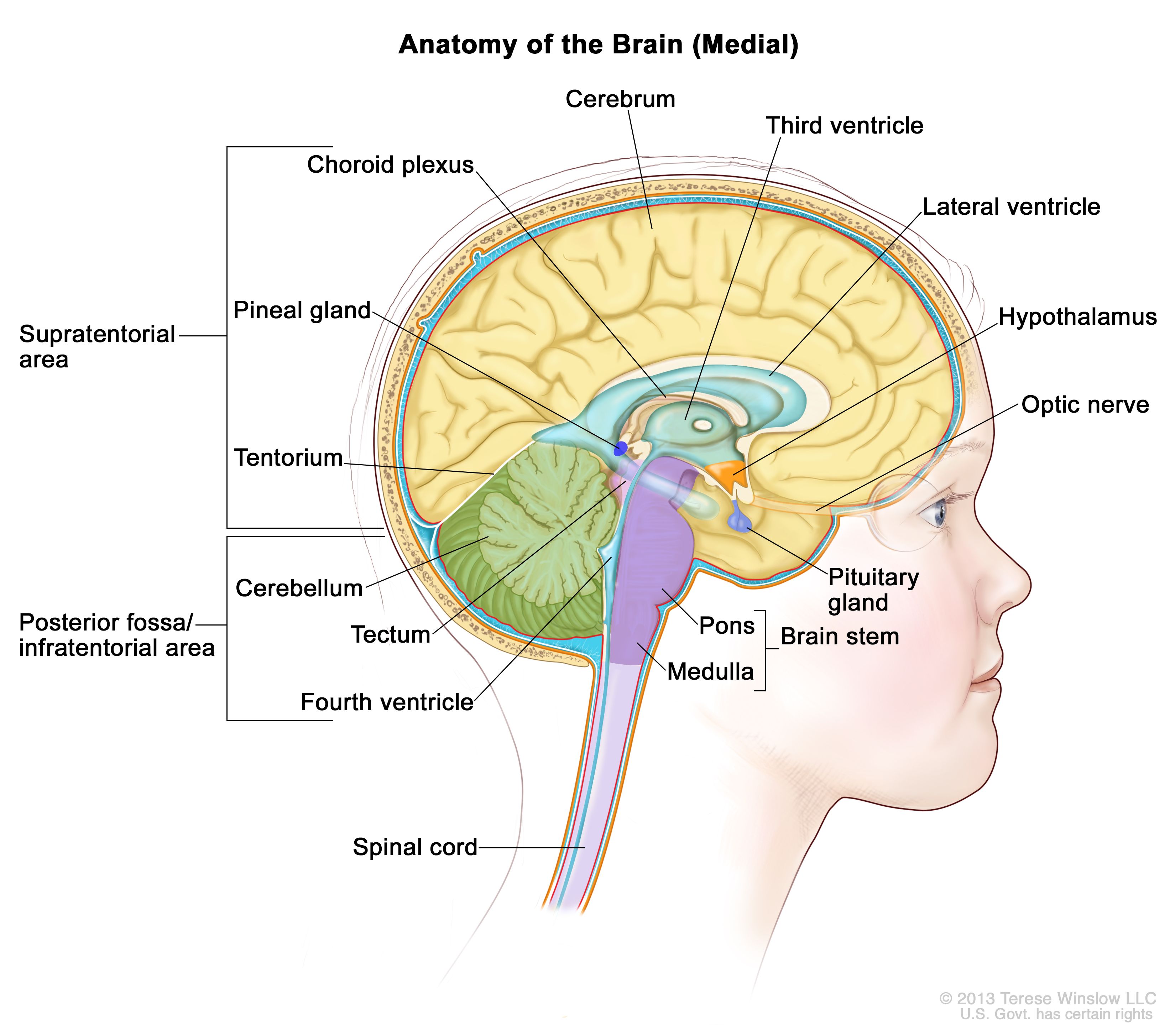
When these cells undergo genetic mutations that cause them to divide uncontrollably, an astrocytoma forms. Because astrocytes are found throughout the brain and spinal cord, these tumors can appear in various locations, including the cerebellum, the cerebrum, the brainstem, or the spinal column. Their behavior—how fast they grow and how likely they are to spread—depends heavily on their genetic profile and histological grade.
In 2026, the medical community views astrocytomas not just as physical masses, but as complex biological entities defined by their molecular signatures. This shift from looking at cells under a microscope to analyzing their DNA has revolutionized how patients are diagnosed and treated.
The 2026 Classification System: WHO Grade and Molecular Markers
The World Health Organization (WHO) classification system is the gold standard for defining astrocytomas. Historically, tumors were graded solely on how "angry" or abnormal the cells looked under a microscope. Today, molecular markers—specific genetic mutations—are the primary drivers of classification.
Low-Grade Astrocytomas (Grades 1 and 2)
Grade 1: Pilocytic Astrocytoma These are typically slow-growing and well-defined tumors. They are most common in children and young adults. Because they often stay in one place and do not infiltrate surrounding tissue as aggressively as higher grades, they are frequently considered "benign" or curable through surgical resection alone.
Grade 2: Diffuse Astrocytoma While still considered low-grade, these tumors are "diffuse," meaning they have finger-like projections that invade neighboring healthy brain tissue. This makes complete surgical removal difficult. As of 2026, a Grade 2 diagnosis requires careful monitoring, as these tumors have a known tendency to evolve into higher-grade malignancies over time.
High-Grade Astrocytomas (Grades 3 and 4)
Grade 3: Anaplastic Astrocytoma These are malignant tumors that grow more rapidly than Grade 2. They exhibit increased cellular density and more frequent cell division (mitosis). Treatment usually requires a combination of surgery, radiation, and chemotherapy.
Grade 4: Astrocytoma, IDH-mutant In previous decades, all Grade 4 astrocytic tumors were often lumped under the term "Glioblastoma." However, modern neuro-oncology differentiates between Glioblastoma (IDH-wildtype) and Grade 4 Astrocytoma (IDH-mutant). The presence of the IDH mutation typically suggests a slightly better prognosis and a different therapeutic path compared to the more aggressive IDH-wildtype glioblastoma. These tumors are characterized by necrosis (cell death within the tumor) and rapid blood vessel growth (vascular proliferation).
Common Symptoms and Clinical Presentation
Symptoms of an astrocytoma are rarely unique to the tumor itself; rather, they are caused by the tumor’s location, its size, and the pressure it exerts on the surrounding brain (increased intracranial pressure).
- Seizures: Often the first sign of an astrocytoma, especially in low-grade tumors. New-onset seizures in adults always warrant neuroimaging.
- Headaches: These are typically worse in the morning and may be accompanied by nausea or vomiting. They result from the buildup of pressure within the skull.
- Cognitive and Personality Changes: Tumors in the frontal lobe may cause changes in mood, judgment, or social behavior.
- Focal Neurological Deficits: Depending on the tumor's location, a patient may experience weakness on one side of the body (hemiparesis), vision loss, speech difficulties (aphasia), or balance issues.
- Memory Loss: Common when tumors involve the temporal lobes or deep brain structures.
The progression of symptoms can be a clue to the tumor grade. Low-grade astrocytomas may cause subtle symptoms that persist for years, while high-grade tumors typically cause a rapid decline in function over weeks or months.
Diagnostic Protocols and Molecular Testing
As of 2026, the diagnostic journey begins with advanced neuroimaging but culminates in a sophisticated molecular analysis.
Advanced Imaging
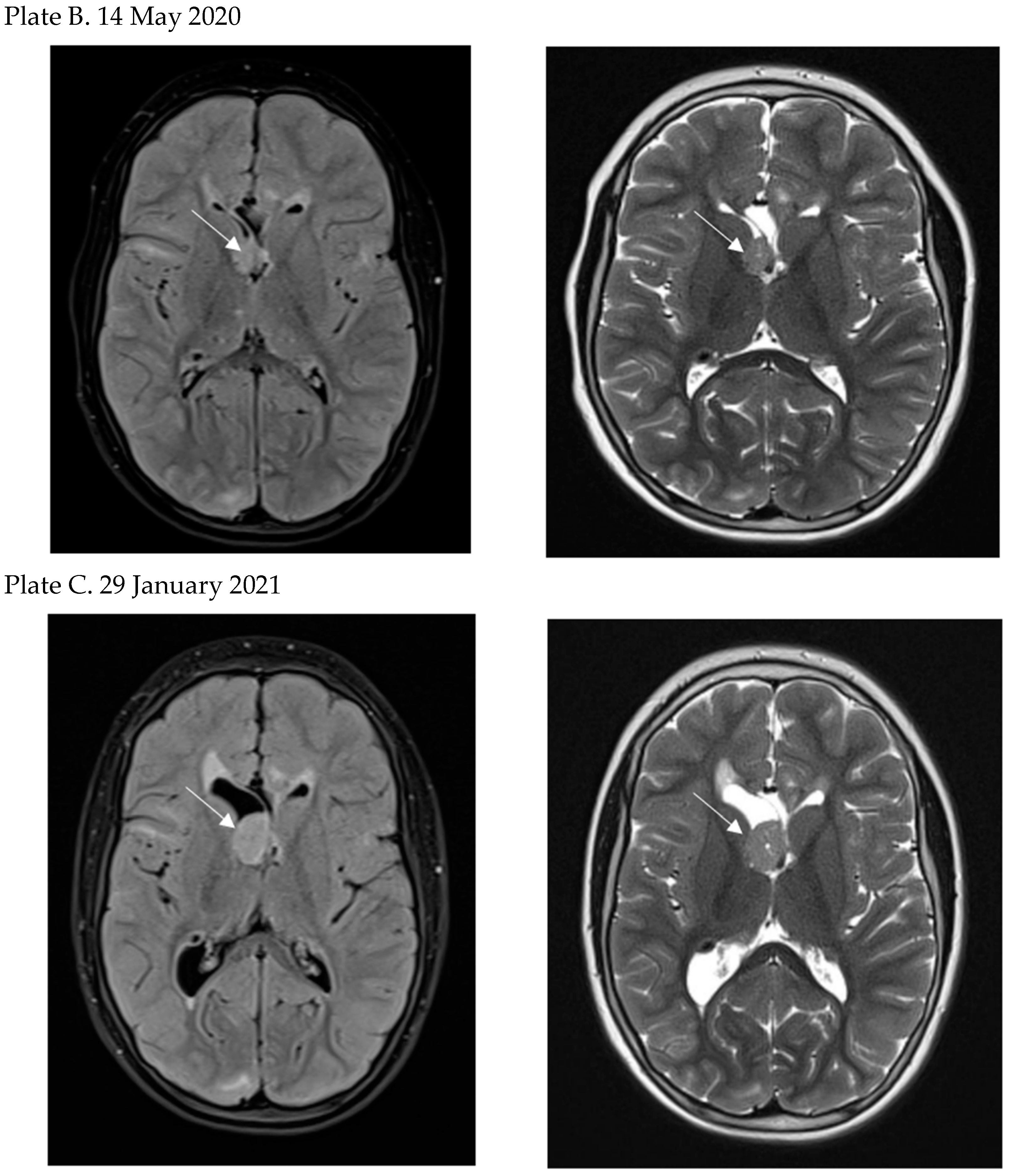
Magnetic Resonance Imaging (MRI) with contrast is the primary tool. Doctors look for "enhancement," where the tumor "lights up" after an injection of gadolinium. Enhancement often suggests a higher grade because it indicates a breakdown in the blood-brain barrier. Functional MRI (fMRI) and Diffusion Tensor Imaging (DTI) are used to map critical areas of the brain—like speech and motor centers—to plan safe surgical routes.
Biopsy and Resection
A definitive diagnosis requires tissue. This is obtained either through a stereotactic needle biopsy (a small hole in the skull) or a craniotomy (surgical removal of the tumor).
Molecular Profiling
Once tissue is removed, pathologists perform several key tests:
- IDH1 and IDH2 Mutation Status: This is the most critical marker for determining the tumor's lineage.
- MGMT Promoter Methylation: Patients with a "methylated" MGMT promoter typically respond much better to chemotherapy (Temozolomide).
- 1p/19q Codeletion: This test helps distinguish astrocytomas from oligodendrogliomas.
- CDKN2A/B Deletion: In 2026, the loss of this gene is recognized as a marker for high-grade behavior, even if the cells look low-grade under a microscope.
Comparison of Astrocytoma Grades and Characteristics
The following table summarizes the typical clinical profiles for astrocytomas based on the current 2026 standards of care and classification.
| Feature | Grade 1 (Pilocytic) | Grade 2 (Diffuse) | Grade 3 (Anaplastic) | Grade 4 (IDH-mutant) |
|---|---|---|---|---|
| Typical Age | Children/Young Adults | 20s to 40s | 30s to 50s | 40s to 60s |
| Growth Rate | Very Slow | Slow to Moderate | Fast | Very Fast |
| Common Location | Cerebellum / Optic Nerve | Cerebrum (Hemispheres) | Cerebrum | Cerebrum |
| Primary Goal | Complete Resection/Cure | Control/Delay Progression | Aggressive Debulking | Maximal Safe Resection |
| Common Markers | BRAF fusion/mutation | IDH mutation, ATRX loss | IDH mutation, p53 | IDH mut, CDKN2A loss |
| Treatment Mode | Surgery (often sufficient) | Surgery + Observation/RT | Surgery + RT + Chemo | Surgery + RT + Chemo |
Treatment Options and Multimodal Management
Management of an astrocytoma is rarely a single-step process. In 2026, the "Standard of Care" involves a multidisciplinary team including neurosurgeons, neuro-oncologists, and radiation oncologists.
Surgical Intervention
The primary goal is Maximal Safe Resection. Surgeons aim to remove as much of the tumor as possible without damaging areas of the brain responsible for movement, speech, or cognition. In some cases, "Intraoperative MRI" or "Fluorescence-guided surgery" (using a dye that makes tumor cells glow) is used to improve precision. For tumors in deep or sensitive areas, Laser Interstitial Thermal Therapy (LITT)—a minimally invasive heat treatment—may be an option.
Radiation Therapy
Radiation uses high-energy beams to kill remaining tumor cells. Standard treatment usually involves six weeks of daily sessions. Advanced techniques like Proton Therapy or Stereotactic Radiosurgery (SRS) are used to focus radiation more tightly on the tumor, sparing healthy brain tissue and reducing long-term cognitive side effects.
Chemotherapy and Targeted Therapy
Temozolomide (TMZ) remains the backbone of chemotherapy for high-grade astrocytomas. It is an oral pill taken during and after radiation. For patients with specific genetic profiles, newer targeted therapies may be used to inhibit specific growth pathways within the tumor cells.
Tumor Treating Fields (TTFields)
For high-grade astrocytomas, TTFields (Optune) may be prescribed. This involves wearing a device on the scalp that creates electric fields, which disrupt the ability of cancer cells to divide. In 2026, this is a common adjunct to chemotherapy for Grade 4 tumors.
Prognosis and Survival Rates
Prognosis is highly individualized. While statistics provide a general idea, they do not predict the outcome for a specific person. Key factors influencing survival include:
- Age: Younger patients generally have better outcomes.
- Extent of Resection: Removing more than 90% of the visible tumor significantly improves the outlook.
- Molecular Profile: IDH-mutant tumors have a much more favorable prognosis than IDH-wildtype tumors.
- Performance Status: The patient’s overall health and level of independence at the time of diagnosis.
Low-grade astrocytomas (Grade 1 and 2) often allow for many years or even decades of high-quality life. High-grade astrocytomas remain challenging, but advancements in immunotherapy and personalized vaccines in clinical trials are continuously extending life expectancy.
Coping with a Diagnosis: Next Actions
Receiving an astrocytoma diagnosis is life-altering. Beyond the immediate medical treatments, several steps are vital for long-term management:
- Seek a Second Opinion: Ensure the diagnosis and molecular testing have been reviewed at a high-volume brain tumor center or NCI-designated cancer center.
- Genetic Counseling: While most astrocytomas are sporadic (not inherited), understanding the genetics of the tumor can help in choosing the right clinical trials.
- Neuro-Rehabilitation: If the tumor or surgery has affected speech, motor skills, or memory, start physical or occupational therapy early to maximize recovery.
- Palliative and Supportive Care: This is not just for end-of-life; it focuses on managing symptoms, reducing stress, and improving quality of life from day one.
Frequently Asked Questions
Is an astrocytoma a brain cancer?
The term "cancer" usually refers to malignant tumors (Grades 3 and 4). Grade 1 astrocytomas are generally considered benign but are still serious because they are located within the skull. Grade 2 tumors are often referred to as "low-grade gliomas" and occupy a middle ground—they are not technically "cancerous" in their initial state but are treated as potentially malignant due to their invasive nature.
Can astrocytomas be cured?
Grade 1 (Pilocytic) astrocytomas are often considered curable if they can be completely removed by a surgeon. Higher-grade astrocytomas (Grades 2, 3, and 4) are currently considered treatable but not typically "curable" in the traditional sense, as microscopic cells often remain in the brain. The goal of treatment for these grades is to manage the disease as a chronic condition.
How is an astrocytoma different from a glioblastoma?
As of the 2021/2026 classification standards, a "Glioblastoma" is specifically an IDH-wildtype Grade 4 tumor. An "Astrocytoma, IDH-mutant, Grade 4" is a different biological entity. While both are aggressive, the IDH-mutant version usually occurs in younger patients and has a statistically better response to treatment and a longer survival rate.
Is an astrocytoma hereditary?
In the vast majority of cases, no. Most astrocytomas are the result of acquired genetic mutations that happen by chance throughout a person's life. However, they can be associated with rare genetic syndromes like Neurofibromatosis type 1 (NF1), Li-Fraumeni syndrome, or Turcot syndrome.
What is the most important factor in the success of treatment?
The most significant factors are the molecular markers (specifically IDH status) and the extent of surgical resection. A tumor that is fully resectable and carries the IDH mutation generally has the most optimistic outlook among the primary gliomas.