Advanced dental implants encompass specialized surgical techniques, computer-guided planning systems, and immediate-loading protocols designed to replace missing teeth efficiently, even in patients with severe bone loss or complex medical histories. These interventions utilize high-tech imaging, digital impressions, and biocompatible materials like titanium and zirconia to restore function and aesthetics with high predictability.
Understanding the Evolution of Dental Implantology
Traditional tooth replacement protocols often required multiple separate surgeries, extensive healing periods spanning six to twelve months, and numerous restorative phases. Modern implant dentistry has evolved significantly through the integration of digital technology, advanced biomaterials, and refined surgical philosophies. Today, clinicians utilize three-dimensional diagnostic imaging and computer-aided design software to evaluate bone density, nerve pathways, and sinus cavities with sub-millimeter precision before making a single incision.
This shift from analog estimation to digital precision has transformed recovery times and expanded eligibility. Patients who were previously told they lacked sufficient bone structure for traditional fixtures can now often benefit from specialized grafting methods or zygomatic designs that anchor into the cheekbone rather than the upper jaw. By shifting the focus toward minimally invasive placement and immediate functional loading, contemporary practices minimize post-operative discomfort and reduce the overall duration of the treatment lifecycle.
Core Technologies Driving Advanced Procedures
The success of modern tooth replacement relies heavily on specialized hardware and software working in tandem. Cone beam computed tomography (CBCT) scanners capture high-resolution, three-dimensional views of the maxilla and mandible, offering far superior diagnostic clarity compared to traditional two-dimensional dental radiographs.
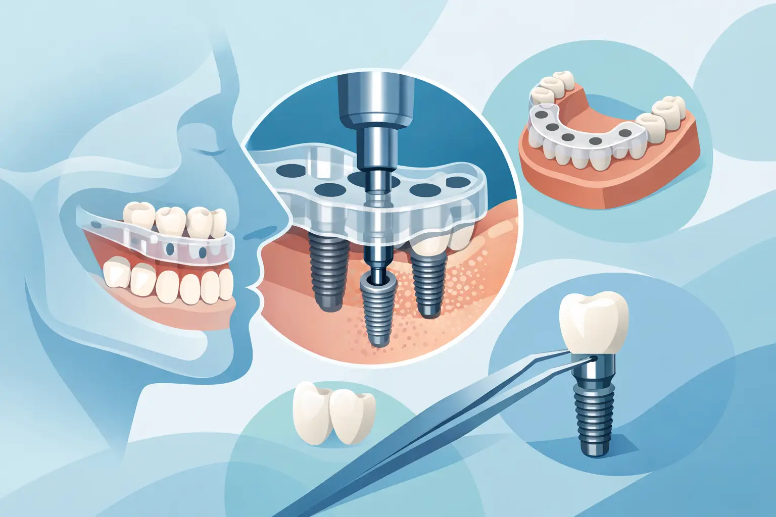
Once the clinician acquires the CBCT data, it is merged with digital surface scans of the patient's dentition using specialized CAD/CAM software. This allows the surgical team to perform a virtual surgery, testing various implant sizes, angles, and depths on a computer screen. From this digital plan, laboratories can print custom surgical guides. These physical templates snap securely over the teeth or gums during the procedure, forcing the surgical drills to follow the exact trajectory mapped out during the virtual planning phase.
Key Technological Components
- Cone Beam Computed Tomography (CBCT): Provides a 360-degree, three-dimensional rendering of bone architecture, blood vessel paths, and nerve locations.
- Computer-Aided Design and Manufacturing (CAD/CAM): Enables the digital design of custom surgical guides, temporary restorations, and permanent prosthetics.
- Dynamic Navigation Systems: Utilize real-time optical tracking to guide surgical instruments with the precision of GPS mapping during live operations.
- Intraoral Scanners: Replace messy physical impression trays with rapid digital optical captures of the oral cavity.
Specialized Surgical Techniques for Complex Cases
Not all patients present with ideal anatomical conditions. Periodontal disease, long-term tooth absence, trauma, and congenital conditions frequently result in localized bone resorption. Advanced dental implantology addresses these structural deficiencies through specialized surgical approaches that reconstruct the lost foundation.
Zygomatic Implants
For patients with severe maxillary bone atrophy who wish to avoid extensive, multi-stage bone grafting, zygomatic options offer a viable alternative. Instead of anchoring into the resorbed upper jawbone, these fixtures are significantly longer and are anchored securely into the dense zygomatic bone (the cheekbone). This technique provides immediate stability, allowing clinicians to mount a full arch of teeth on the same day as the surgery, even when traditional upper jaw bone density is virtually non-existent.
All-on-4 and Full-Arch Rehabilitation
The All-on-4 protocol—and its multi-implant variants—allows an entire upper or lower arch of teeth to be supported by as few as four strategically placed fixtures. The posterior fixtures are typically tilted at a 30 to 45-degree angle to maximize contact with available bone and avoid critical anatomical structures like the inferior alveolar nerve or maxillary sinuses. This angular placement creates a wide tripod effect, distributing bite forces evenly across the restoration and frequently eliminating the need for bone grafting.
Immediate Placement and Loading
In traditional workflows, an extraction is performed, the site is left to heal for several months, an implant is placed, and another healing period follows before the final crown is attached. Immediate placement allows the clinician to insert the fixture into the fresh extraction socket during the same appointment. Under favorable primary stability conditions, immediate loading protocols permit the attachment of a temporary restoration on the very same day, preserving soft-tissue architecture and eliminating the period of living without visible teeth.
Comparison of Standard Versus Advanced Approaches
| Parameter | Standard Dental Implants | Advanced Dental Implants |
|---|---|---|
| Diagnostic Imaging | 2D panoramic or periapical radiographs | 3D Cone Beam Computed Tomography (CBCT) |
| Surgical Placement | Freehand placement based on visual inspection | Computer-guided templates or dynamic navigation |
| Bone Grafting Needs | Often requires separate, staged grafting phases | Utilizes tilt, zygomatic, or immediate techniques to bypass grafting |
| Treatment Timeline | 6 to 18 months with multiple distinct phases | Often streamlined to single-visit or expedited multi-phase protocols |
| Anatomical Limitations | High rejection rate in severe bone loss cases | Adaptable to compromised or severely resorbed jaws |
Clinical Prerequisites and Patient Evaluation
Determining candidacy for advanced procedures requires a comprehensive medical and dental evaluation. While modern techniques accommodate many complex cases, systemic health factors play a critical role in osseointegration—the biological process where living bone cells fuse directly to the surface of the titanium or zirconia fixture.
Clinicians evaluate systemic conditions such as uncontrolled diabetes, autoimmune disorders, and active oncological treatments, as these can impair healing and increase the risk of fixture failure. Active periodontal disease must be treated and stabilized prior to any surgical intervention to prevent peri-implantitis, an inflammatory condition that can destroy the supporting bone around the restoration.
Furthermore, lifestyle factors such as tobacco use significantly impact success rates. Nicotine restricts blood flow to the gingiva and bone, slowing the crucial cellular regeneration required for successful osseointegration. Smokers are counselled regarding these elevated risks and are often required to participate in strict cessation programs prior to surgery.
Step-by-Step Treatment Lifecycle
Understanding the sequence of advanced procedures helps patients navigate their treatment journey with realistic expectations. While timelines vary based on individual anatomical requirements, a typical computer-guided, full-arch or complex single-fixture workflow follows a structured path.
- Comprehensive Diagnostics: Acquisition of CBCT scans, digital surface impressions, and a thorough medical history review to map out the anatomical landscape.
- Virtual Planning: Digital simulation of the surgery to select optimal fixture diameters, lengths, and insertion trajectories, culminating in the fabrication of a custom surgical guide.
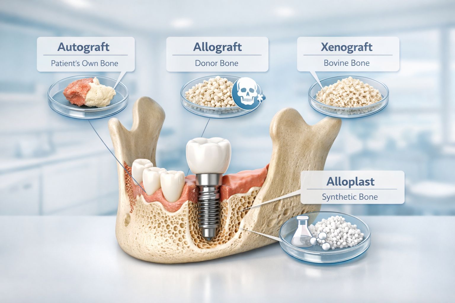
- Site Preparation and Extraction: Removal of any failing teeth, minor debridement of infected tissue, and execution of localized bone preservation or grafting if necessary.
- Guided Placement: Insertion of the fixtures through the surgical guide, ensuring precise alignment and achieving optimal primary mechanical stability.
- Provisionalization: Attachment of a lightweight, non-functional or functional temporary restoration, depending on the immediate loading criteria met during surgery.
- Osseointegration Period: A biological healing phase lasting several months, during which bone tissue remodels and locks the fixture securely in place.
- Final Restoration: Replacement of the provisional prosthesis with a permanent, highly durable prosthetic crafted from monolithic zirconia or porcelain-fused-to-metal.
Potential Complications and Maintenance Protocols
Advanced surgical procedures carry inherent risks that patients must understand. While success rates generally exceed 95 percent, biological and mechanical complications can occur. Biological complications include early failure of osseointegration (often due to infection or excessive early loading) and late-onset peri-implantitis, a progressive inflammatory disease characterized by bone loss around an integrated fixture.
Mechanical complications involve wear, chipping, or fracture of the prosthetic components, as well as the loosening of abutment screws over time due to parafunctional habits like bruxism (teeth grinding). Patients who grind their teeth are typically prescribed custom night guards to protect both the restoration and the opposing natural dentition.
Long-term success depends heavily on rigorous daily hygiene and professional maintenance. Because artificial teeth cannot decay, many patients assume they require less maintenance. However, plaque and calculus accumulate on implants just as they do on natural teeth, making diligent brushing, specialized flossing tools, and regular professional cleanings mandatory to ensure the longevity of the investment.
Frequently Asked Questions
Are advanced dental implants painful?
Surgical placement is performed under local anesthesia, intravenous sedation, or general anesthesia, ensuring the patient feels no discomfort during the procedure. Post-operative discomfort is generally managed effectively with prescribed or over-the-counter analgesics, and most patients report that the recovery is much easier than anticipated.
How long do advanced dental implants last?
With proper oral hygiene, routine professional maintenance, and favorable systemic health, the titanium or zirconia fixture can last a lifetime. The visible prosthetic crown, bridge, or denture attached to the top may experience normal wear over 10 to 15 years and may eventually require repair or replacement.
Can smokers receive advanced dental implants?
Yes, but smoking significantly increases the risk of implant failure due to compromised blood flow and delayed healing. Clinicians evaluate each smoker on an individual basis and strongly recommend cessation before and after the surgical phase to maximize the probability of successful osseointegration.
What is the difference between standard and guided surgery?
Standard surgery relies on the surgeon's visual estimation and freehand tool manipulation during the operation. Guided surgery utilizes 3D computer software and custom physical templates to direct drills and fixtures to exact pre-planned locations, reducing surgical time and minimizing tissue trauma.
How do zygomatic options differ from standard ones?
Standard fixtures anchor directly into the alveolar bone of the upper or lower jaw. Zygomatic fixtures are substantially longer and bypass the upper jaw entirely, anchoring deep into the zygomatic bone (cheekbone) to provide immediate stability for patients with severe upper jaw bone loss.
Recommended Next Actions
If you are considering advanced dental implant treatment, schedule a comprehensive consultation with a qualified periodontist, oral and maxillofacial surgeon, or restorative dentist with specialized training in digital implantology. Request a complete diagnostic workup involving a CBCT scan to evaluate your bone volume and structural anatomy accurately. Ensure your provider discusses all available options—including bone grafting, guided surgery, and immediate-loading protocols—tailored specifically to your clinical presentation and long-term health goals.